どうも、Beyond the Pixelです。外科手術の進化は目覚ましく、より複雑な手技が可能になる一方で、手術中の神経損傷リスクへの配慮はこれまで以上に重要になっています。米国神経生理学的モニタリング学会(ASNM)が最近発表した「術中筋電図(EMG)モニタリングに関する最新のポジションステートメント」は、この課題に対する明確な指針を示しています。これは、2005年の以前の声明を更新し、手術中に末梢運動軸索の機能的状態を評価し、対象者の神経学的完全性を保護するためのEMGの推奨される使用法を詳細に解説しています。
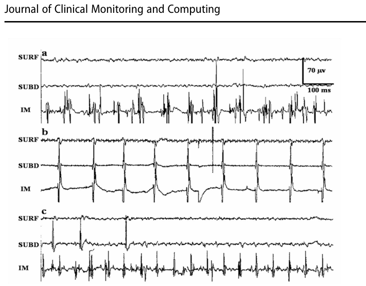
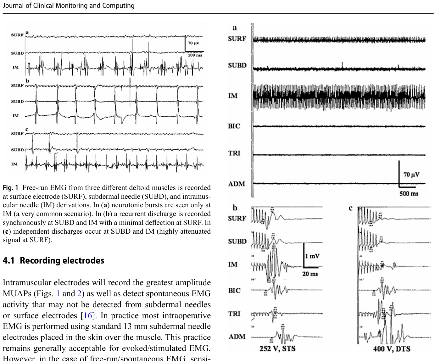
Figure 1. Fig. 1 Free-run EMG from three different deltoid muscles is recorded at surface electrode (SURF), subdermal needle (SUBD), and intramus cular needle (IM) derivations. In (a) neurotonic bursts are seen only at IM (a very common scenario). In (b) a recurrent discharge is recorded synchronously at SUBD and IM with a minimal deflection at SURF. In (c) independent discharges occur at SUBD and IM (highly attenuated signal at SURF).解説: デルタ筋から記録されたフリーランEMGの例を示します。筋内電極(IM)では、皮下電極(SUBD)や表面電極(SURF)では検出されにくい神経緊張性バーストがはっきりと見られます。神経筋活動の記録において、電極の配置と種類がいかに重要かを示しています。Figure 2. Fig. 2 Surgeon alerted after 20–30 + Hz right deltoid A-train discharge, detected mainly at IM during free-run EMG (a). Small deflections seen at SURF & SUBD. Pre-discharge TCE MEP (b) reveals good signal acquisition at deltoid sites (especially IM). After A-train, TCE MEP amplitude reduced > 90% (despite higher stimulation voltage and dou ble train stimulation (DTS) (c). Post-operatively, patient suffered com plete long-standing right deltoid paralysis. SURF, SUBD, IM = right deltoid surface, subdermal needle, and intramuscular needle deriva tions respectively. BIC = biceps brachii, TRI = triceps, ADM = abduc tor digiti minimi right IM needle derivations. STS = single train stimulation.解説: 右デルタ筋のフリーランEMGで検出された20-30+HzのA-train放電と、その後の経頭蓋電気刺激運動誘発電位(TCE MEP)の変化を示しています。この放電後、TCE MEPの振幅が90%以上減少し、術後に右デルタ筋の完全麻痺に至ったケースです。
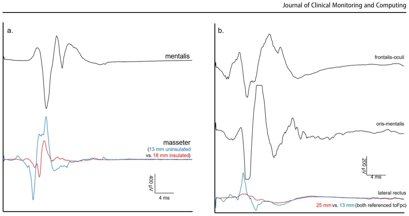
Figure 3. Fig. 3 Impact of an insulated shaft electrode on decreasing unwanted triggered EMG from overlapping cranial myotomes. (a) Triggered EMG responses after cathodal monopolar stimulation of CN 7 at 1.0 mA with a 50 µs pulse were recorded using 13 mm uninsulated needle electrodes (1.5 cm spacing) from mentalis (black trace) and masse ter (blue trace). A 25 mm insulated shaft electrode with only 6 mm of the tip uninsulated was placed at the same location for masseter recording (red trace). All responses were recorded at 400 µV/Div using a passband filter of 30 − 10,000 Hz. The unwanted contamina tion from CN 7-innervated muscles (presumably platysma, possibly others like buccinator) shows up as a larger waveform in the uninsu lated (blue) pair of electrodes than the insulated pair (red). (b) Trig gered EMG responses were recorded using 13 mm uninsulated needle electrodes from frontalis-oculi (top black trace), oris-mentalis (bottom解説: 絶縁されたシャフト電極が、隣接する頭蓋神経筋からの不要な誘発性EMGを低減する効果を示しています。絶縁されていない電極(青い線)では顔面神経刺激による大きな波形が記録されるのに対し、絶縁された電極(赤い線)ではそれが抑制されています。これにより、特定の筋肉からの活動をより選択的に記録できることが示唆されます。
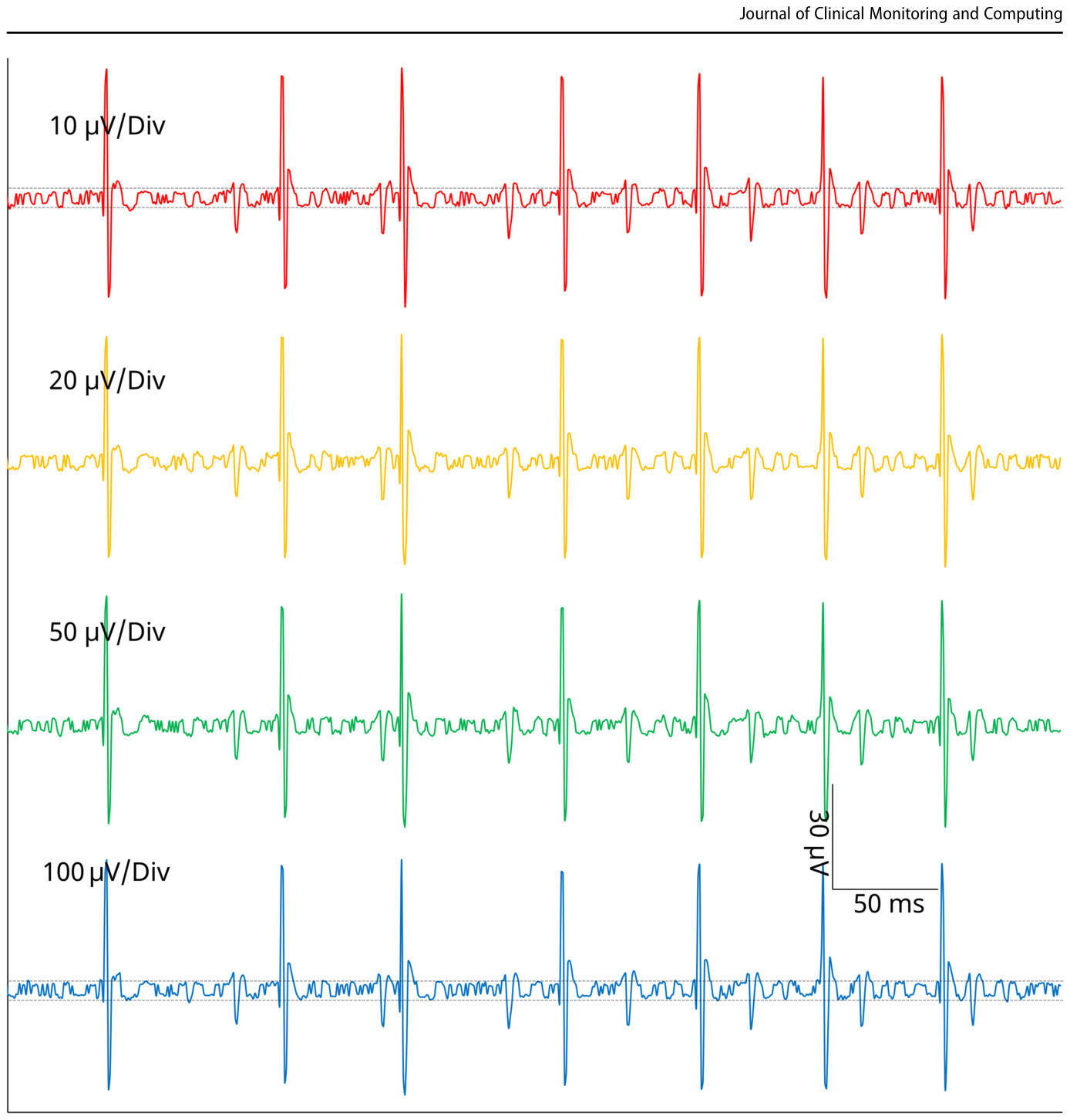
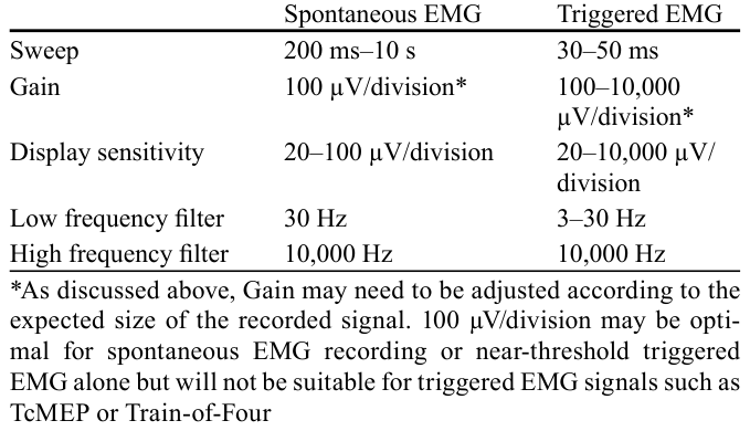
Figure 4. Fig. 4 Impact of changing analog-to-digital converter (ADC) ranges without gaining vertical resolution. Spontaneous EMG from mentalis was recorded at four “gain” settings (10, 20, 50, and 100 µV/Div) with intraoperative neurophysiologic monitoring equipment which allows changes in “gain” that do not use different levels of amplification but only different ADC ranges. Intrinsic amplifier noise (dotted lines ≈ 5 µV) is larger than digitized steps of the 16-bit ADC, which are 0.002解説: アナログ-デジタル変換器(ADC)のレンジを変更しても、垂直分解能が必ずしも向上しない影響を示しています。異なる「ゲイン」設定(10、20、50、100 µV/Div)での自発性EMG記録を比較しており、アンプの固有ノイズがデジタル化されたステップよりも大きいため、一部のゲイン設定ではダイナミックレンジが犠牲になるだけで、波形分解能は向上しないことを示しています。
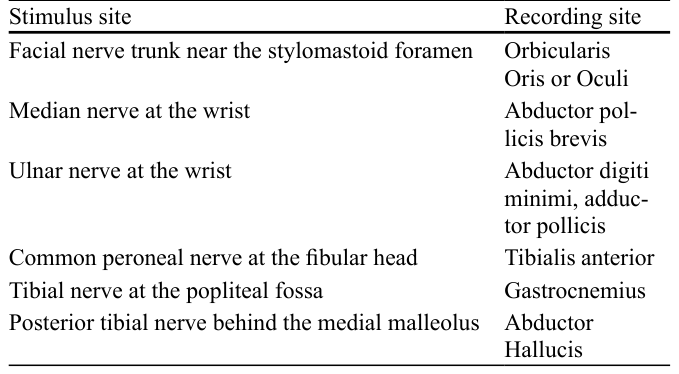
Table 3. Table 3 Common stimulus and recording sites for Train-of-Four (TOF) testing Stimulus site Recording site Facial nerve trunk near the stylomastoid foramen Orbicularis Oris or Oculi Median nerve at the wrist Abductor pol licis brevis Ulnar nerve at the wrist Abductor digiti minimi, adduc tor pollicis Common peroneal nerve at the fibular head Tibialis anterior Tibial nerve at the popliteal fossa Gastrocnemius Posterior tibial nerve behind the medial malleolus Abductor Hallucis解説: 4連反応(TOF)テストの一般的な刺激部位と記録部位をまとめた表です。顔面神経、正中神経、尺骨神経、総腓骨神経、脛骨神経など、各神経に対応する刺激部位と、そこから筋電図を記録する筋肉が示されています。
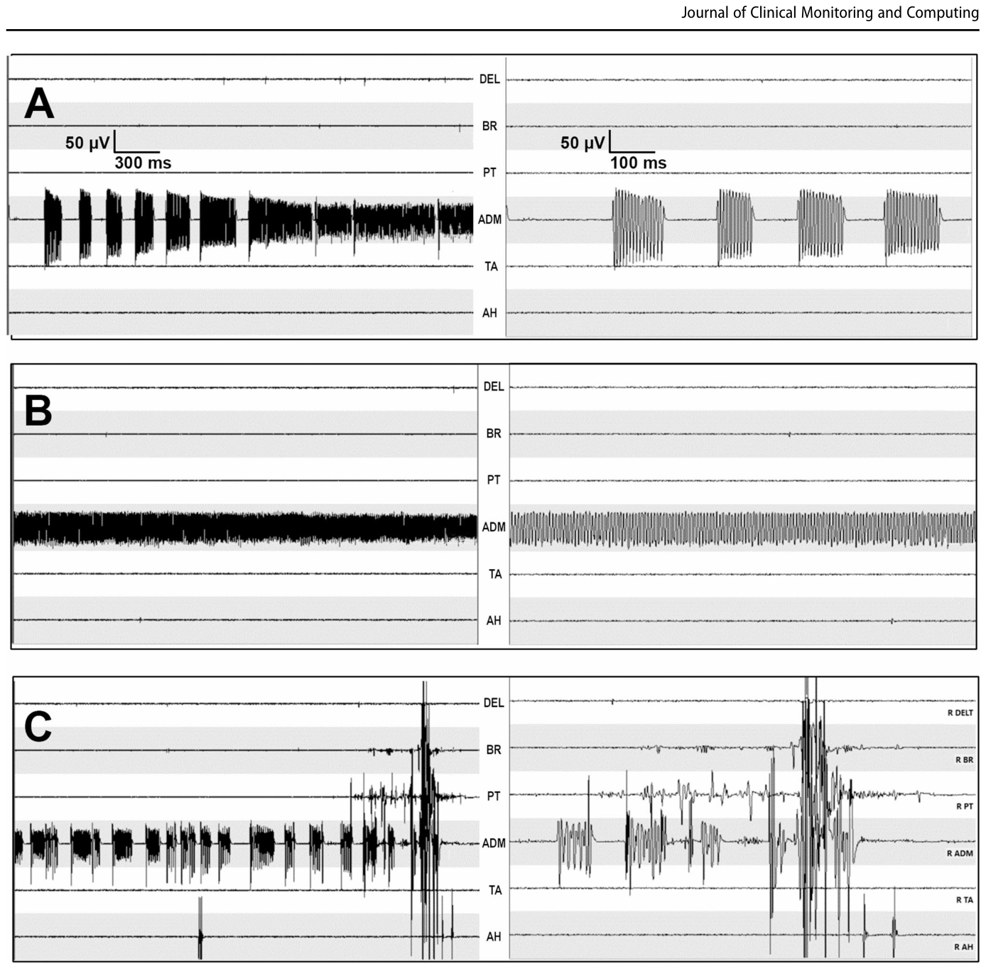
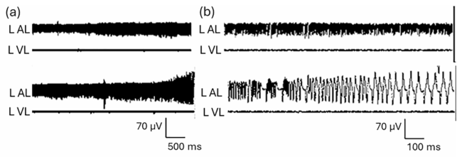
Figure 6. Fig. 6 Middle-aged female with neck pain and C7–T1 spondylolisthe sis undergoes decompression/fusion. Left = 3, right = 1 s time sweep. (a) During active decompression at C7-T1, bursts of 140 Hz A-trains are reported; (b) The bursting A-trains evolve into a continuous 140 + Hz train activity; (c) A-train salvos re-appear, convert to non-解説: C7-T1脊椎すべり症の対象者における術中EMG記録の例です。減圧術中に140HzのA-trainバーストが報告され、その後連続的な活動に移行し、最終的に右腕の特定の筋肉(R ADM)のTcMEP消失と術後麻痺につながったケースを示しています。Figure 8. Fig. 8 Abrupt A-train is generated after prone positioning in a patient with L2 –L3 pseudoarthrosis and an unstable spine. The peak fre quency is 200 Hz, but A-train frequency may vary dramatically as in the lower trace of (b). The trace duration is 5 s in (a), 1 s in (b). Upper traces of (a) & (b) record the early A-train epoch, lower traces the later evolution解説: L2-L3偽関節と不安定な脊椎を持つ対象者が腹臥位になった際に発生した急なA-train放電を示しています。ピーク周波数は200Hzですが、A-trainの周波数は大きく変動する可能性があります。
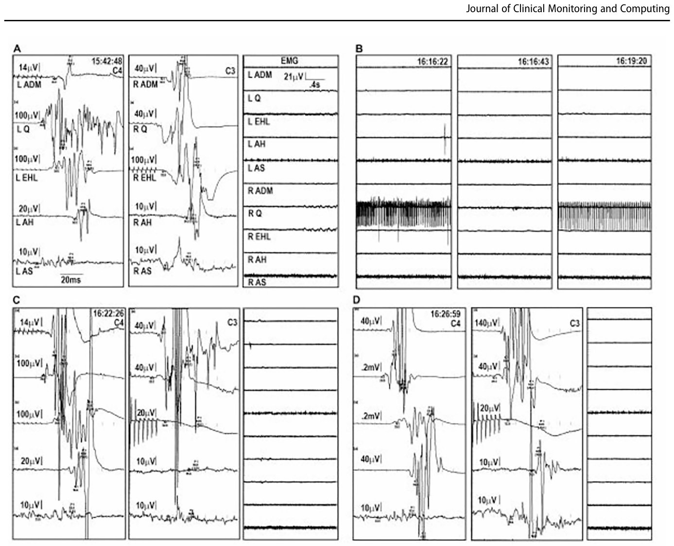
Figure 11. Fig. 11 20 yo F with dysesthesiae in both legs and T11–T12 HNP under goes transforaminal T11–T12 thoracic interbody fusion. (a) Before rod compression, C4 and C3 TcMEP and EMG are normal; (b) Rods are compressed with immediate R Q SED (16:16:22). SED subsides after release of rod compression (16:16:43) but returns (16:19:20) when rods are re-compressed. (c) TcMEP lost at R EHL, AH with maintained rod compression (16:22:26); (d) TcMEP recovering when kyphosis reduc解説: T11-T12椎間孔経路椎体間固定術中のTcMEPとEMG記録を示しています。ロッド圧迫後、即座に右四頭筋に上位セグメント発生EMG放電(SED)が発生し、これは神経学的変化の早期指標となり得ます。
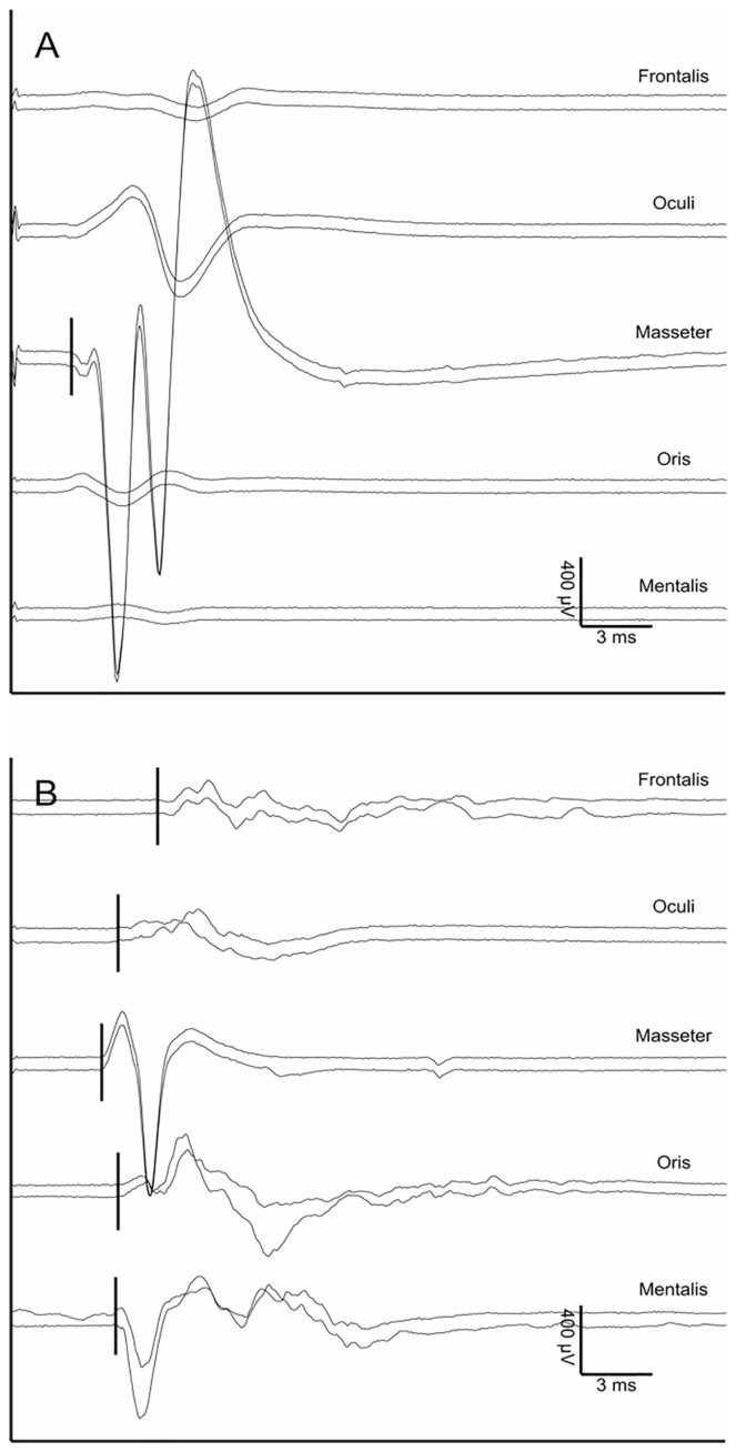
Figure 12. Fig. 12 Onset latency facilitates differentiation of cranial nerve trig gered EMG responses. Bipolar needle electrode recordings from cra nial nerve V and VII innervated muscles show compound motor action potentials (CMAPs) after stimulation of cranial nerves V (a) or VII (b). The proximity of muscles innervated by these two nerves results in volume conduction “crosstalk” between the channels. However, the onset latency of masseter CMAPs are shorter than those originating from facial innervated muscles解説: 頭蓋神経の誘発性EMG応答の「発症潜時」が、異なる頭蓋神経を識別するのに役立つことを示しています。例えば、顔面神経(VII)と三叉神経(V)刺激後のマスセター筋(咬筋)の応答潜時の違いを通じて、神経を特定できます。
Figure 13. Fig. 13 Lateral spread response in patient with hemifacial spasm. With stimulation of the zygomatic branch of the facial nerve, an expected early onset latency direct CMAP occurs in orbicularis oculi (a), whereas a longer latency “lateral spread response” (LSR) occurs in
Figure 14
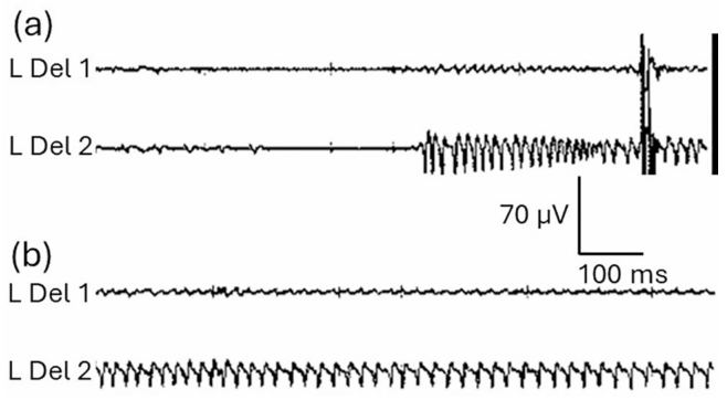
Figure 14. Fig. 14 In order to avoid inadequate sampling, two channels of left deltoid EMG are recorded during anterior cervical discectomy C4-C5 (L Del 1 & 2). In (a), the abrupt onset of an A-train is clearly seen at L Del 2; in (b), the L Del 2 A-train continues to exhibit typically low amplitude and variable frequency features
Figure 15
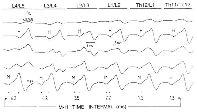
Figure 15. Fig. 15 Soleus responses following dorsal midline lumbar stimulation at six sites from L4-L5 to T11-T12. Low and high stimulating intensity evoked indirect (H / PRM reflex) and direct (M) responses respectively at all sites. The H-to-M interval shortens as stimulation moves crani ally away from the emergence of S1-S2 soleus roots. Minimum H-M values are seen at T11-T12 and T12-L1.
Figure 17
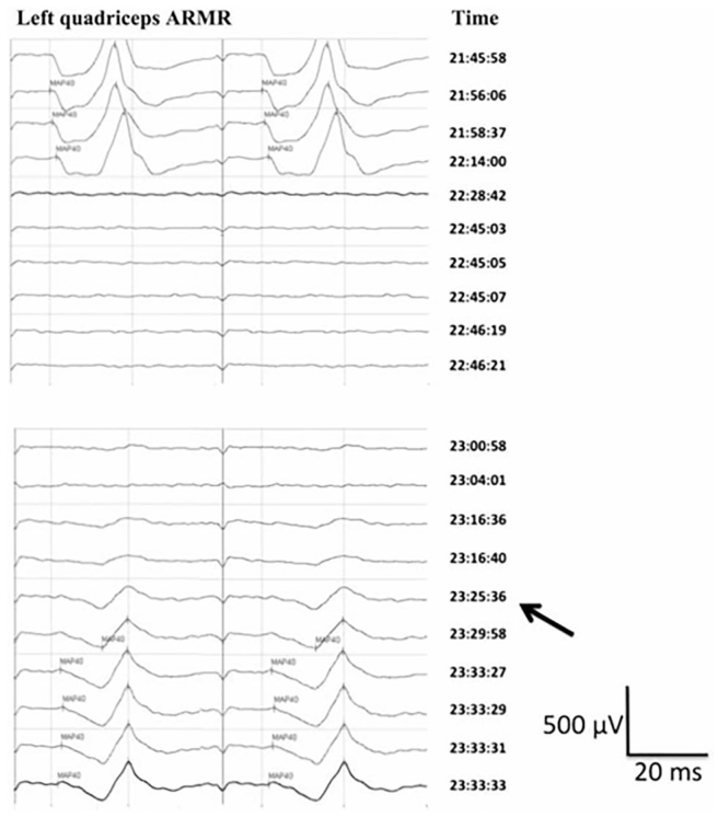
Figure 17. Fig. 17 Sudden isolated loss of ARMR in quadriceps muscle while a Hohmann retractor was compressing the femoral nerve (22:28:42). The response recovered post-retractor removal. Arrow = start of response recovery. Recovery from mild post-operative quadriceps palsy within 48 h. ARMR, Anterior root muscle response = TaCMAP in this review.


コメントを残す