どうも、Beyond the Pixelです。遠位型ミオパチー(DM)や筋原線維性ミオパチー(MFM)は、手足の遠位部(指先や足先に近い部分)の筋肉に影響を及ぼす遺伝性の病気で、その遺伝的多様性ゆえに診断が非常に難しいことで知られています。これまでの遺伝子検査では診断に至らないケースも多く、より効率的な診断方法が求められていました。近年、筋肉MRIは、これらの疾患における筋肉の変化パターンを捉える上で重要なツールとなっており、特定の疾患に特徴的な画像バイオマーカー(生体指標となる画像情報)を特定するのに役立っています。さらに、人工知能(AI)の一つである機械学習技術を組み合わせることで、診断プロセスをさらに進化させる可能性が示されています。この研究は、大規模な多施設共同研究のデータを用いて、これらの希少な筋疾患における筋肉MRIパターンを詳細に解析し、その診断性能を定量化すること、そして機械学習を活用した新しい画像ベースの分類フレームワークを開発することを目的としています。
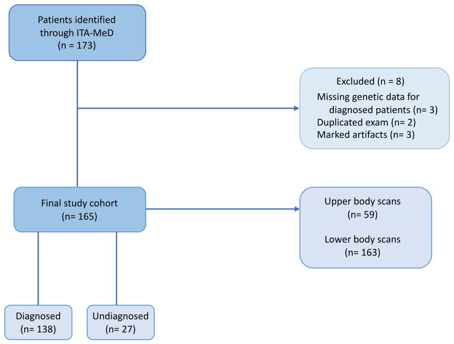
Figure 1. Fig. 1 Flowchart with an overview of the study cohort解説: 本研究の対象者選定プロセスを示しています。当初173名から、除外基準を満たした8名を除き、最終的に165名の対象者が研究に含まれました。このうち138名は遺伝子診断が確定しており、27名は未診断でした。取得されたスキャンは、上半身が59件、下半身が163件でした。
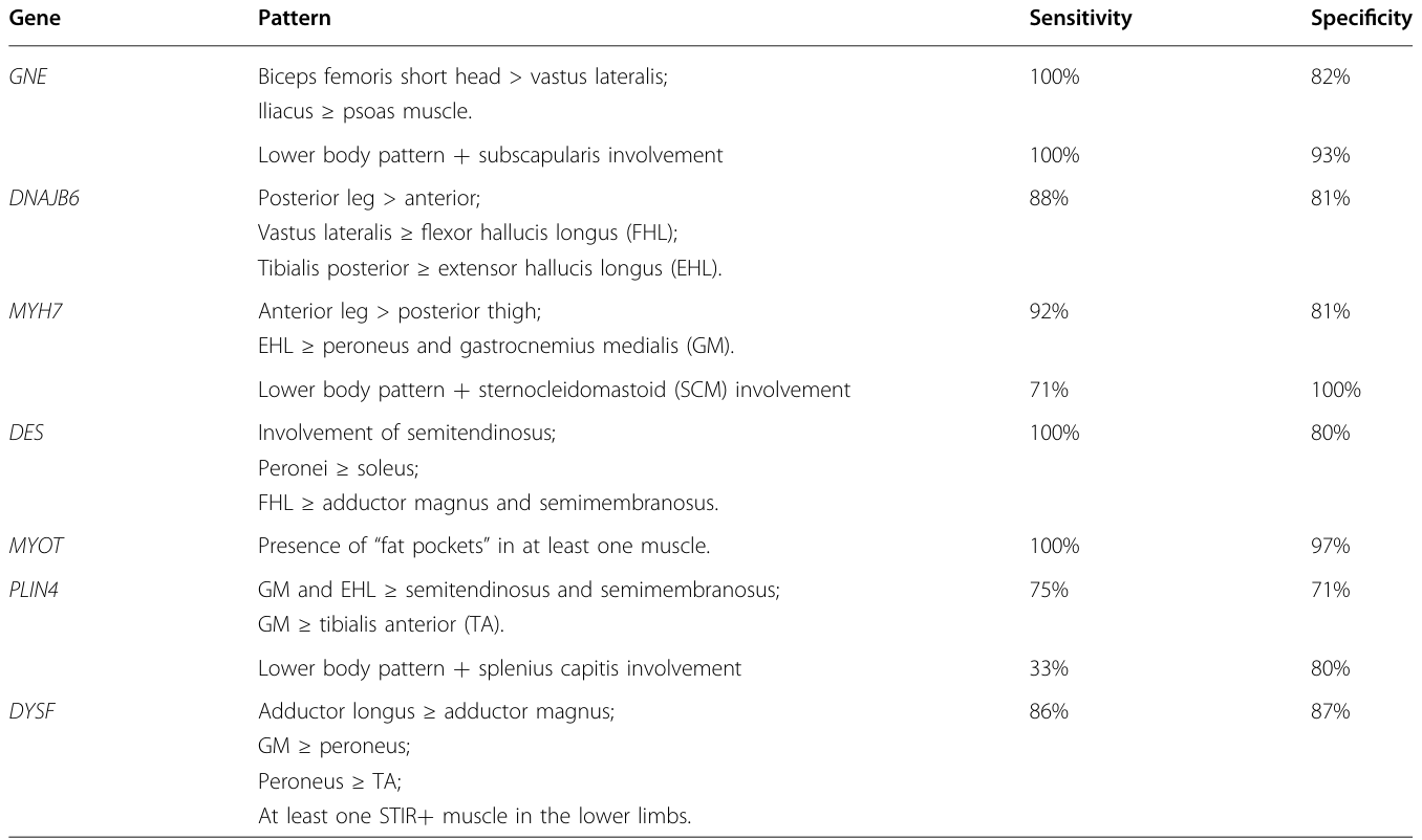
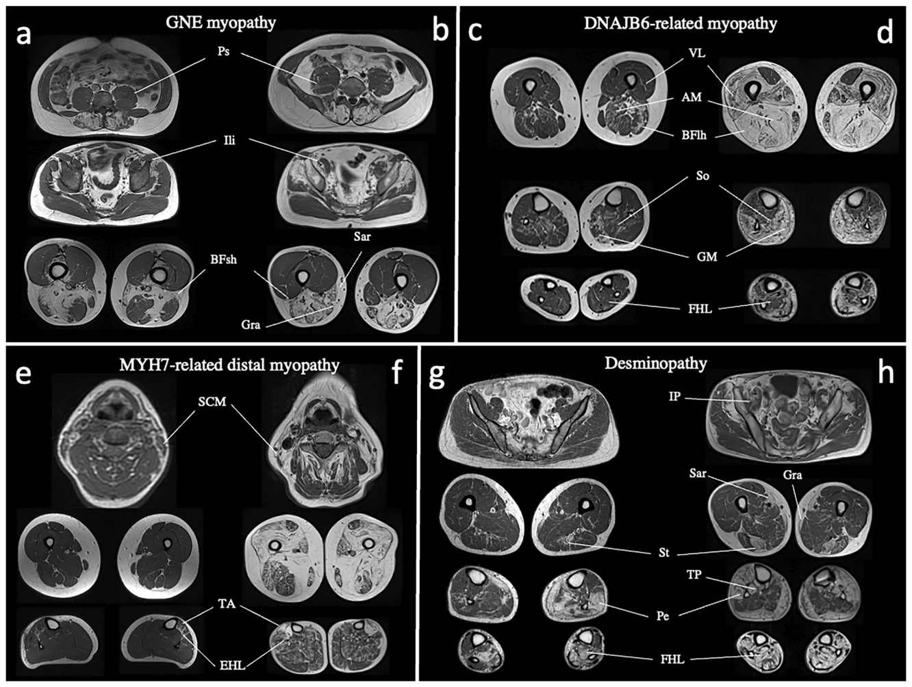
Figure 2. Fig. 2 Patterns of muscle involvement in GNE myopathy, DNAJB6-, MYH7- related myopathies, and desminopathy. GNE myopathy (a): predominant fatty replacement of iliacus (Ili) over the preserved and hypertrophic psoas muscle (Ps) in the pelvis, and complete fatty replacement of BFsh in the thigh in a 36-year-old patient; b the same pattern can be visualised in a 33-year-old patient, along with the complete sparing of vasti muscles and asymmetric involvement of sartorius (Sar) and gracilis muscles (Gra). DNAJB6-related myopathy (c, d): soleus (So) and GM are the most consistently involved muscles together with adductor magnus (AM) and biceps femoris long head (BFlh), while anterior leg muscles are relatively spared; vastus lateralis (VL) is at least as affected as FHL. The pattern can be recognised both in a 44-year-old patient with mild, predominantly proximal MFM (c) and in a 52-year-old patient with a distal myopathy (DM) with myofibrillar pathology phenotype (DM-MP) with more severe disease (d). MYH7-related DM (e, f): EHL and TA are the earliest affected muscles in the lower limbs in a 37-year-old woman; at this stage, mild SCM muscle involvement is already evident in the upper body (e); thigh muscles are severely affected in a 57-year-old woman with more advanced disease, while anterior leg muscles are still more involved than those in the posterior thigh and leg (f). Desminopathy (g, h): FHL, peroneus (Pe) and semitendinosus (St) muscles are invariably involved; sartorius (Sa) and gracilis (Gra) are frequently affected as well (g, h), both in a 40-year-old man with mild disease (g) and in a 42- year-old man (h) with greater severity. With disease progression (h), both anterior and posterior leg muscles, including tibialis posterior (TP), may become fatty replaced, together with iliopsoas (IP) in the pelvis解説: GNEミオパチー、DNAJB6関連ミオパチー、MYH7関連ミオパチー、デスミノパチーにおける特徴的な筋肉関与パターンをMRI画像で示しています。例えば、GNEミオパチーでは腸骨筋の脂肪変性と大腿二頭筋短頭の関与が、MYH7関連ミオパチーでは前下腿の伸筋群(EHLやTA)の早期関与が見られます。
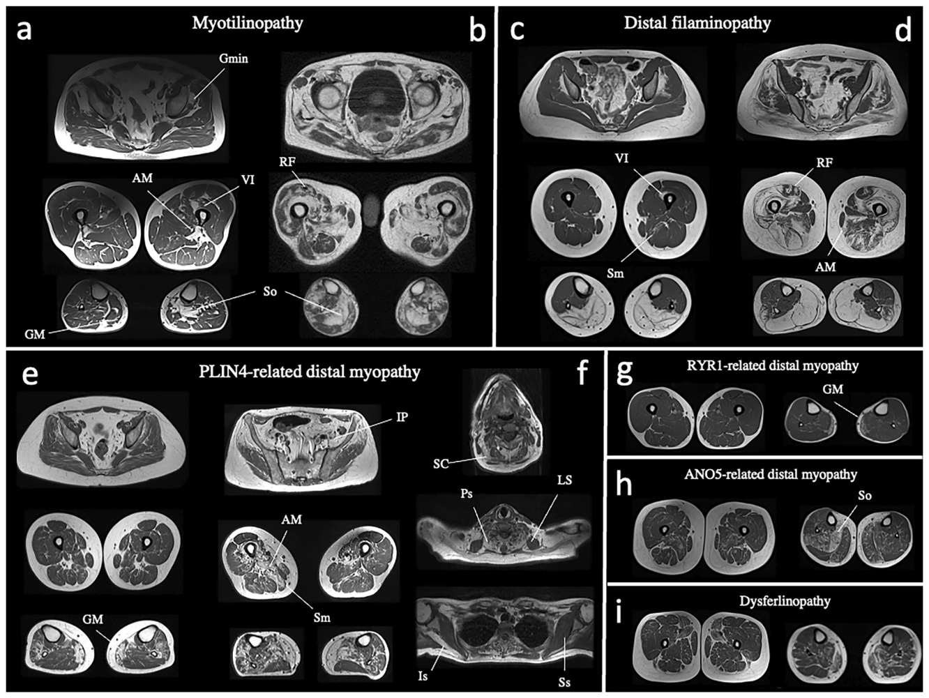
Figure 3. Fig. 3 Patterns of muscle involvement in myotilinopathy, distal filaminopathy, PLIN4-, RYR1-, and ANO5-related distal myopathies, and dysferlinopathy. Myotilinopathy: a, b Since the early disease stages, «fat pockets» can be visualised in the most frequently involved muscles in the disease, such as adductor magnus (AM), vastus intermedius (VI), soleus (So), GM, and gluteus minimus (Gmin). These areas tend to become confluent as muscle pathology progresses (b). Asymmetry is another common feature of myotilinopathies, as seen in the soleus muscles (So) of a 38-year-old man (a) and in the rectus femoris (RF) of a 72-year-old man (b). Distal filaminopathy (c, d): calf involvement in a 29-year-old patient, along with early and asymmetric affection of semimembranosus (Sm) and vastus intermedius (VI) (c). More advanced muscle pathology in a 60-year-old relative of the same patient, with peculiar involvement of some muscles in the subfascial (vasti, adductor magnus—AM) or myotendinous junction (rectus femoris —RF) regions (d). PLIN4-related DM (e, f): anterolateral leg muscles and GM are the earliest and most consistently involved muscles (e), as can be seen in a 36-year-old woman. With disease progression, semimembranosus (Sm), adductor magnus (AM), and vasti muscles become involved, often asymmetrically, along with iliopsoas (IP) (f). Asymmetry can also be seen in the upper body of the same patient, where a peculiar involvement of splenius capitis (SC) among the neck extensors can be found, together with cervical paraspinal (Ps) and infraspinatus muscles (Is). Levator scapulae (LS) and subscapularis (Ss) are selectively spared (f). Calf muscle involvement in RYR1-, ANO5- related DM and dysferlinopathy (g–i): in RYR1-related DM (g), isolated GM involvement can be a feature, while it is more often associated with concomitant variable, asymmetric involvement of soleus (So) and adductor muscles in ANO5-related DM (h) and distal dysferlinopathy (i)解説: ミオチリノパチー、遠位型フィラミノパチー、PLIN4関連DM、RYR1関連DM、ANO5関連DM、ディスフェルリノパチーにおける筋肉の関与パターンをMRI画像で示しています。ミオチリノパチーでは「脂肪ポケット」と呼ばれる多発性の脂肪変性が特徴的です。PLIN4関連DMでは前外側下腿筋と腓腹筋内側頭が早期に侵されます。
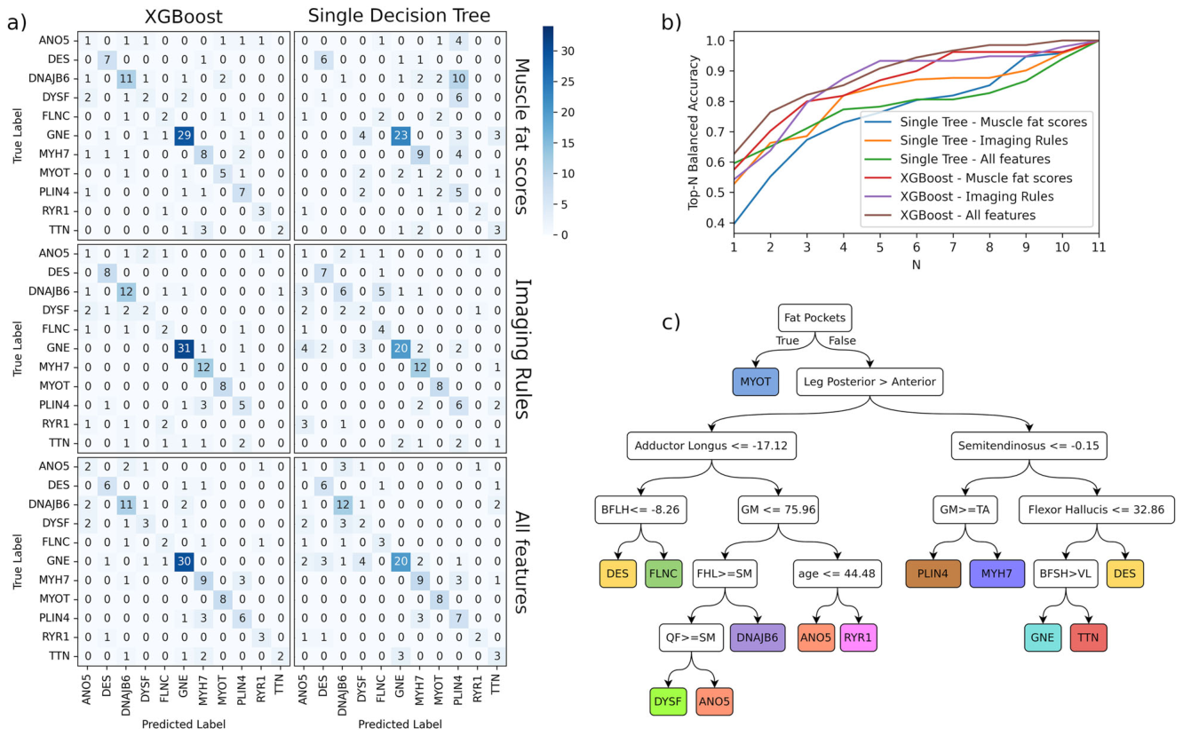
Figure 4. Fig. 4 Machine learning analysis and output. Confusion matrices for XGBoost and Single Decision Tree models using the three different datasets (T1- scores, imaging rules and all features combined) (a). Each matrix shows the classification results for genetic entities; rows represent the true genetic diagnosis (true label) and columns the predicted one (predicted label). Values in the diagonal indicate correct predictions: colour intensity reflects the number of correctly or incorrectly classified cases, with darker shades indicating higher counts. Top-N Balanced accuracy for XGBoost and Single Decision Tree model (b). The Top-N accuracy indicates whether the correct genetic diagnosis falls within the top N predictions ranked by model probability. Across all values of N, models using all features (T1 scores + imaging rules) generally outperformed those using only one feature set, with the XGBoost model achieving the highest overall accuracy. Single Decision Tree model (c). Each node poses a binary question, proceeding to the left if the answer is yes and to the right if the answer is no until a terminal node is reached, indicating the predicted outcome. The presence of “fat pockets” was chosen as the most informative feature, allowing identification of all patients with myotilinopathy. In the absence of this feature, the anterior or posterior predominance of leg involvement splits the tree into two major branches, highlighting the relevance of regional distribution patterns. Downstream, specific muscle involvement thresholds help differentiate between diseases解説: 機械学習モデル(XGBoostと単一決定木)の分析結果と出力が示されています。混同行列(a)は、各遺伝子疾患の分類結果を示し、対角線上の値が正しい予測を意味します。XGBoostは単一決定木よりも高いバランス精度(b)を達成しています。単一決定木モデル(c)は、診断の意思決定プロセスを視覚的に示し、「脂肪ポケット」の有無がミオチリノパチーの識別に最も重要な特徴として選ばれています。
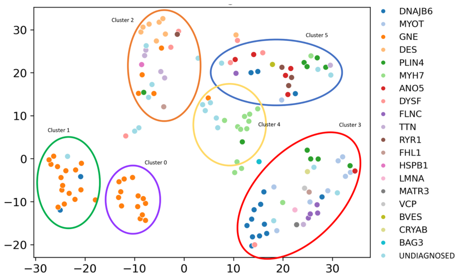
Figure 5. Fig. 5 UMAP-based clustering of muscle MRI scans. Two-dimensional UMAP projection based on z-scores of T1-weighted signal intensity of lower body muscles, capturing patterns of fatty replacement in patients with genetically confirmed and undiagnosed DM/MFM. Six distinct clusters are identified by unsupervised clustering. Coloured dots represent individual patients, coded by the gene harbouring pathogenic variants, while light blue dots denote undiagnosed patients. GNE myopathy patients cluster tightly into two different clusters (0 and 1), mainly defined by differences in disease severity and age at MRI scan, confirming the distinctive imaging phenotype of this cohort of patients. Most of the other clusters are enriched for one or a few specific genetic conditions, such as myopathies caused by DNAJB6, MYOT and FLNC variants for cluster 3, or by MYH7 variants for cluster 4. Patients with variants in DYSF and PLIN4 are more dispersed across multiple clusters. Only a few molecularly diagnosed patients remain unclustered, underscoring the robustness of the UMAP-based model in capturing key radiological patterns解説: 筋肉MRIスキャンのUMAP(均一多様体近似と投影)ベースのクラスタリング結果を示しています。下半身の筋肉におけるT1強調信号強度のzスコアに基づいた二次元UMAP投影で、遺伝子診断確定対象者と未診断DM/MFM対象者の脂肪変性パターンを捉えています。6つの異なるクラスターが特定され、GNEミオパチーの対象者はクラスター0と1に密に集積しています。未診断対象者の多くも、分子診断が確定した対象者のグループ内に位置しています。
コメントを残す