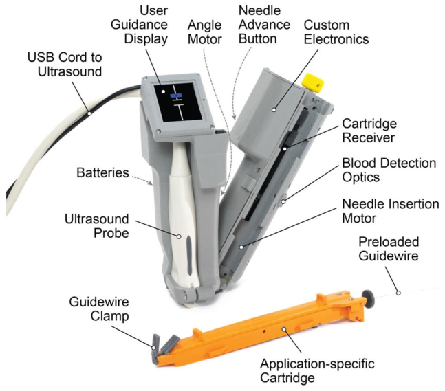
どうも、Beyond the Pixelです。外傷性出血や心停止といった緊急事態において、命を救うためには迅速な血管カニューレ挿入(血管内にカテーテルや針を挿入すること)が不可欠です。しかし、この手技には高い専門知識と経験が求められ、特に資源が限られた環境では熟練した専門家が常にいるとは限りません。本研究で評価されたAI-GUIDE-LLは、このような課題を解決するために開発された画期的なハンドヘルド型の超音波ガイドロボットデバイスです。AIと超音波を組み合わせることで、経験の浅い医療提供者でも大腿血管へのアクセスを可能にすることを目指しています。これまでの研究では、熟練した専門家によるブタモデルでの有効性が示されていましたが、経験の浅い専門家が人体で使用した場合の性能は未知数でした。本研究は、このAI-GUIDE-LLが、超音波ガイド手技の経験が少ない専門家でも、人体のファントム(人体のCTデータから作成された模擬モデル)における大腿血管への針の正確な挿入、および健常なボランティア対象者の大腿血管の特定において、どの程度の性能を発揮するかを評価しました。
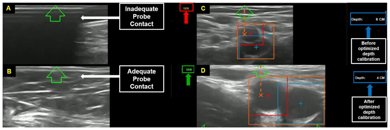
Figure 3. Fig. 3 A Example of inadequate probe-skin contact. The algorithm detects incomplete acoustic coupling (white arrow) and alerts the operator by displaying “Skin” in orange (red arrow). B Example of adequate probe-skin contact. The algorithm confirms appropriate acoustic coupling (white arrow) and signals this to the operator by displaying “Skin” in green (green arrow). Good contact is reflected by uniform echogenicity and clear visualization of superficial tissue layers. C Initial vessel detection and segmentation. The common femoral vessels are identified (orange box) and automatically classified as artery (red box) and vein (blue box). The current imaging depth is 6 cm (green box); the algorithm will subsequently adjust this depth to optimize visualization of the target vessels. D Optimized depth calibration. The AI algorithm has automatically adjusted imaging depth from 6 cm to 4 cm (green box), thus improving visualization of the femoral vessels and providing the operator with clearer spatial orientation to facilitate accurate probe repositioning to center the target artery解説: この図は、プローブと皮膚の接触状態と初期の血管検出および深度校正の例を示しています。Aは不適切なプローブと皮膚の接触の例で、アルゴリズムが不完全な音響カップリングを検出し、オペレーターに警告します。Bは適切なプローブと皮膚の接触の例で、アルゴリズムが適切な音響カップリングを確認し、オペレーターに信号を送ります。Cは初期の血管検出とセグメンテーションの例で、大腿血管が識別され、動脈と静脈として自動的に分類されます。Dは最適化された深度校正の例で、AIアルゴリズムが画像深度を調整し、大腿血管の視覚化を改善している様子が示されています。
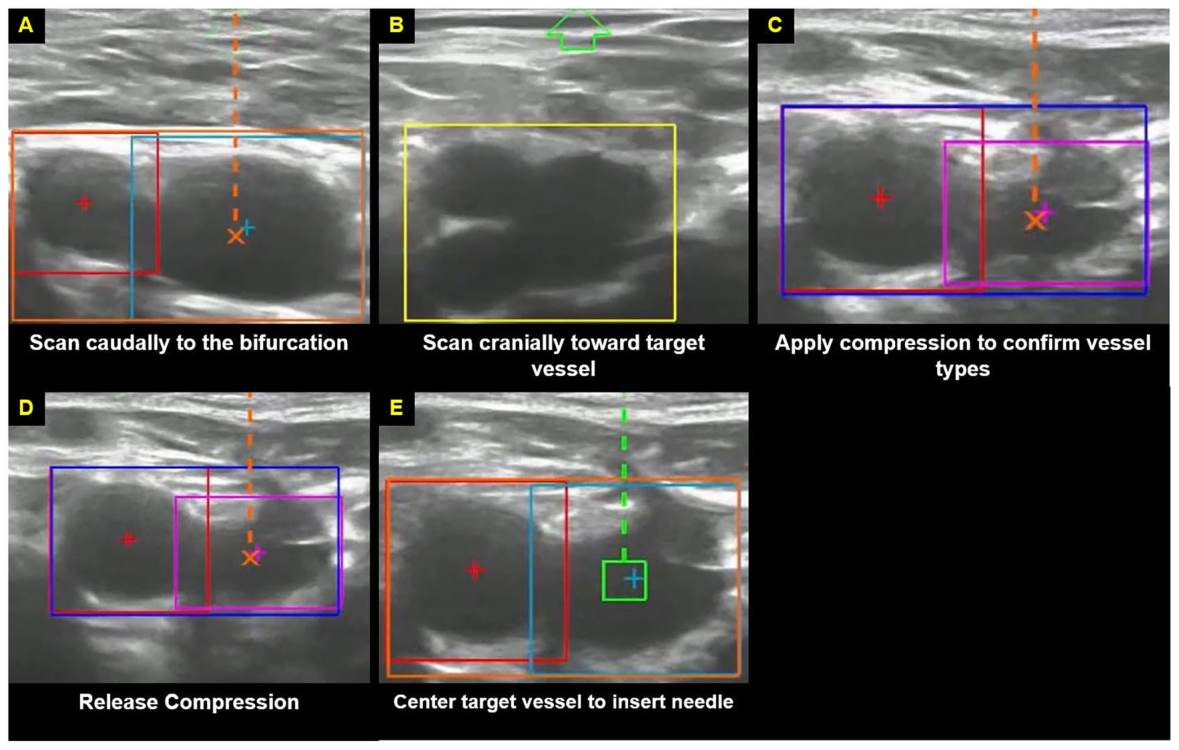
Figure 4. Fig. 4 A (1) Target vessel centering: the operator is instructed to align the probe with the target vein. Proper centering is achieved when the orange X (device reference point) is closely aligned with the blue plus sign (algorithm-identified center of the vein). Once aligned, the operator is guided by written instructions on the device display to “scan caudally towards the bifurcation” to begin stage 2, target vessel confirmation. B (2) Bifurcation identification: the algorithm detects the femoral bifurcation (yellow rectangle). Following successful identification, the operator is instructed to “scan cranially to target vessel” to return to the optimal cannulation site above the bifurcation. C, D (3) Artery-vein differentiation: the operator is prompted to “apply compression to confirm vessel types” to aid in distinguishing the compressible vein from the non-compressible artery. After adequate differentiation is achieved, the operator is instructed to “release compression”. E (4) Final recentering and needle-path configuration: the operator is prompted to perform minor probe adjustments by “Center target vessel to insert needle” to ensure precise recentering of the target vessel. Once alignment is confirmed and the needle trajectory has been automatically configured (green dotted line), the operator is instructed to “push trigger” (on device display) to initiate needle deployment, with the green box indicating the intended entry point within the vein lumen解説: この図は、ターゲット血管の中心合わせから針経路の構成までの一連のワークフローを示しています。Aはターゲット血管の中心合わせの例で、Bは分岐点の識別の例です。CとDは、血管タイプを区別するための圧迫の適用と解除を示しています。Eは、最終的な再中心合わせと針経路の構成の例で、オペレーターが針挿入を開始するよう促される様子が描かれています。
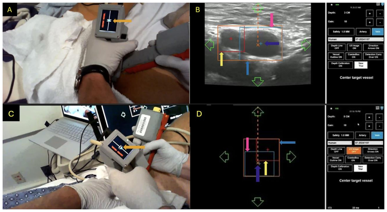
Figure 2. Fig. 2 A In this unblinded human trial, the user places the device onto the scanning volunteer’s right groin with the probe aligned parallel to the inguinal crease. The operator then scans medio-laterally until the target vessel (common femoral vein) is centered and the needle insertion aimpoint is computed. Positioning is guided by aligning the on-screen dot with the crosshair displayed on the device’s LCD (orange arrow). B The user simultaneously receives guidance from the corresponding ultrasound display and software overlay to align the orange X (probe center) with the blue plus sign (common femoral vein center), indicated by the purple arrow. The overlay segments the common femoral artery (yellow arrow), the common femoral vein (pink arrow) within the surrounding box capturing both vessels (blue arrow). C In this blinded human trial, the user places the device on the scanning volunteer’s left groin with the probe aligned parallel to the inguinal crease. The user then scans medio-laterally until the target vessel (common femoral vein) is centered. Positioning is guided by aligning the on-screen dot with the crosshair displayed on the device’s LCD (orange arrow). D The device operator is blinded to the ultrasound display but receives guidance from the corresponding software overlay to align the orange X (probe center) with the blue plus sign (common femoral vein center), indicated by the purple arrow. The overlay segments the common femoral artery (yellow arrow), the common femoral vein (pink arrow) within the surrounding box capturing both vessels (blue arrow)解説: この図は、人体試験におけるデバイスの使用例を示しています。Aでは、非ブラインド条件下でユーザーがデバイスをスキャンボランティア対象者の右鼠径部に置き、プローブを鼠径部のしわと平行に合わせ、ターゲット血管を中心に合わせる様子が示されています。Bは、対応する超音波ディスプレイとソフトウェアオーバーレイからのガイダンスを示しています。Cでは、ブラインド条件下でユーザーがデバイスを左鼠径部に置き、ターゲット血管を中心合わせする様子が示されています。Dは、ブラインド条件下で超音波ディスプレイが見えない状態で、ソフトウェアオーバーレイからガイダンスを受けている様子を示しています。
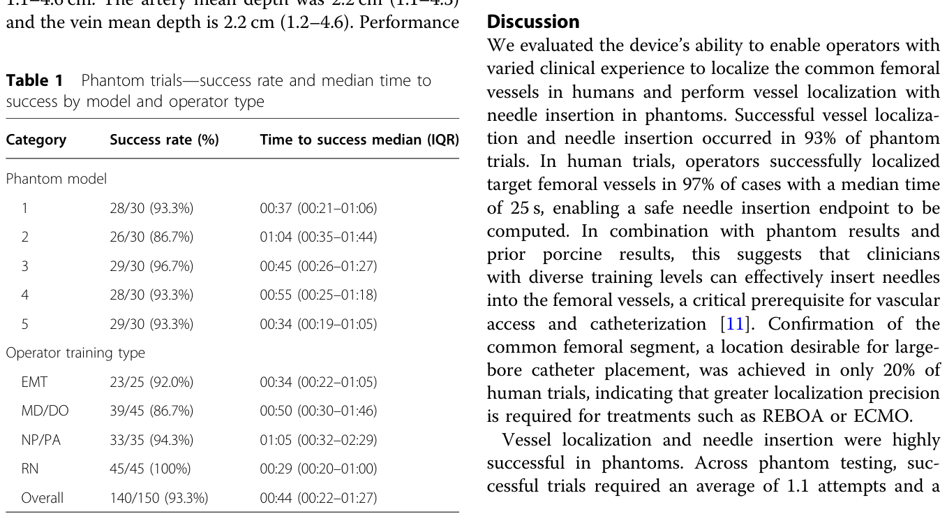
Table 1. Table 1 Phantom trials—success rate and median time to success by model and operator type解説: この表は、ファントム試験におけるファントムモデル別および専門家タイプ別の成功率と、成功までの時間の中央値(四分位範囲)をまとめています。全体の成功率は93.3%で、成功までの中央値時間は44秒でした。登録看護師が100%の成功率を達成しています。
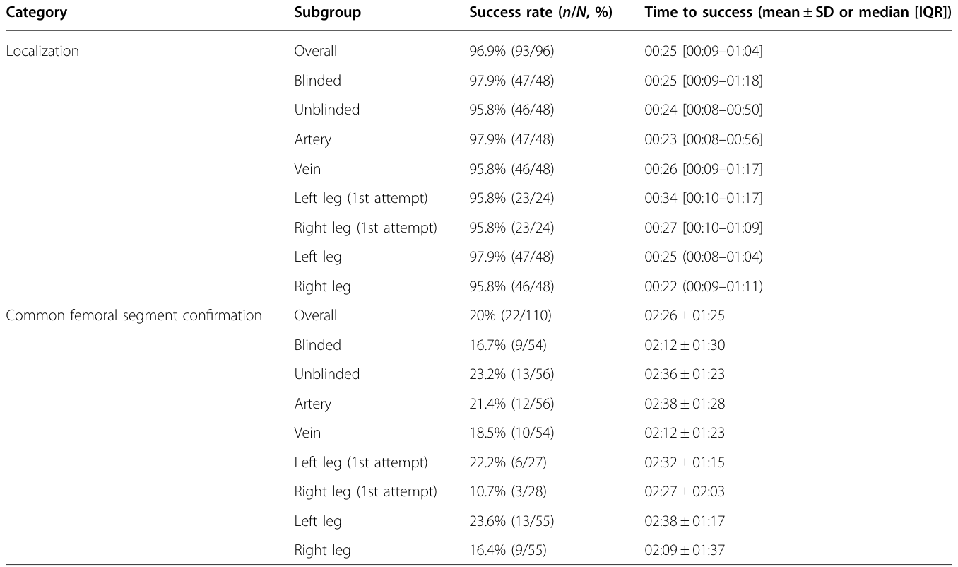
Table 2. Table 2 Human trials—success rate and time to success for vessel localization and target vessel segment confirmation解説: この表は、人体試験における血管特定およびターゲット血管セグメント確認の成功率と、成功までの時間(平均±標準偏差または中央値[四分位範囲])をまとめています。血管特定の全体成功率は96.9%でしたが、大腿共通血管セグメント確認の全体成功率は20%にとどまっています。
Table 3. Table 3 Human trials—success rate and time to success for vessel localization and target vessel segment confirmation (stratified by DO type)解説: この表は、人体試験における血管特定とターゲット血管セグメント確認の成功率および成功までの時間について、デバイスオペレーター(専門家)の種類別に層別化して示しています。上級実践専門家(NP/PA)は、大腿共通血管セグメント確認において31.2%と比較的高い成功率を示しています。
は専門家の種類別、
Table 4. Table 4 Human trials—success rate and time to success for vessel localization and target vessel segment confirmation (stratified by scanning volunteer subgroup)解説: この表は、人体試験における血管特定とターゲット血管セグメント確認の成功率および成功までの時間について、スキャンボランティア対象者のサブグループ(BMI、性別)別に層別化して示しています。BMIが30未満の対象者では大腿共通血管セグメント確認の成功率が24.3%であるのに対し、BMIが30以上の対象者では9.4%と低くなっています。
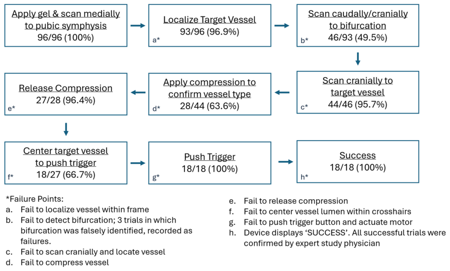
Figure 6. Fig. 6 Human trials full workflow illustrating success rate by step解説: この図は、人体試験の完全なワークフローにおける各ステップごとの成功率を示しています。ゲルの塗布とスキャンから始まり、ターゲット血管の特定、分岐点へのスキャン、血管タイプ確認のための圧迫、圧迫解除、ターゲット血管の中心合わせ、トリガーの押下を経て成功に至るまでの各段階の成功率がパーセンテージで示されています。



コメントを残す