どうも、Beyond the Pixelです。閉塞性睡眠時無呼吸症候群(OSA)は、睡眠中に上気道が繰り返し部分的に、あるいは完全に閉塞することで、酸素飽和度の低下や睡眠分断を伴う疾患です。OSAの管理において、気道閉塞の動態を正確に特徴づけることは極めて重要であり、治療戦略は対象者固有の閉塞パターン特定に大きく依存するようになっています。上気道の磁気共鳴画像(MRI)は、自然睡眠中に解剖学的および機能的評価を行う強力な非侵襲的モダリティとして注目されています。内視鏡技術とは異なり、MRIは気道セグメント全体の管腔狭窄と軟部組織の動きを多層的に評価でき、OSAにおける閉塞が非周期的で空間的に不均一、かつ機械的に複雑であることが多いという点で特に重要です。動的な気道評価は、陽圧呼吸療法に耐えられない対象者にとって効果的な代替手段である舌下神経刺激(HNS)の臨床的関連性が高いと言えます。現在の候補者選定は薬物誘発性睡眠内視鏡(DISE)に依存していますが、新興の証拠は、口蓋帆レベルを超えた閉塞パターン、特に中咽頭の側壁閉塞が治療結果に大きく影響する可能性を示唆しており、生理的な睡眠条件下での包括的な多層的動的評価の必要性を強調しています。しかし、既存の睡眠時MRI法には技術的な限界があります。これまでの研究の多くは、並列イメージングを用いた逐次的な単一スライス収集に依存しており、スライスあたり100~400ミリ秒の時間分解能でした。逐次的な撮像では、スライス間で動きの表現に一貫性がなく、多層的な閉塞の協調的な動きの解釈が困難です。スパイラル収集とビューシェアリング、アンダーサンプリングされた3Dカルテシアン法、そして事前定義された変換正則化(圧縮センシング)を組み合わせた同時多スライスラジアル収集を用いたアプローチは有望でしたが、OSAに特徴的な不規則な呼吸パターン下での時間的ぼやけに敏感であるという課題がありました。
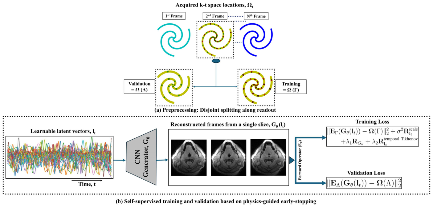
Figure 1. FIGURE 1 | Overview of the proposed self-supervised variational manifold reconstruction framework. (a) For each slice, the acquired k-t spiral measurements are split along the readout direction into disjoint training Ω(Γ) and validation Ω(Λ) subsets. (b) Learnable latent vectors 𝑙𝑡are jointly optimized with a CNN generator 𝐺𝜃that maps 𝑙𝑡to reconstructed image frames for that slice. The generated frames are projected to k-space via the forward operator EΓ, and the training loss enforces data consistency on Ω(Γ) with temporal Tikhonov, generator, and KL regularization terms. The validation loss evaluated on Ω(Λ) is withheld from optimization and used solely to guide early stopping and learning rate reduction. Reconstruction is performed independently for each slice.解説: 提案された自己教師あり変分多様体再構成フレームワークの概要を示しています。(a)各スライスについて、取得されたk-tスパイラル測定値が、読み出し方向に沿って訓練用と検証用に分割されます。(b)学習可能な潜在ベクトルと、その潜在ベクトルを再構成された画像フレームにマッピングするCNNジェネレーターが共同で最適化されます。訓練損失は訓練用データの一貫性を確保し、検証損失は最適化から除外され、早期停止のガイドとしてのみ使用されます。
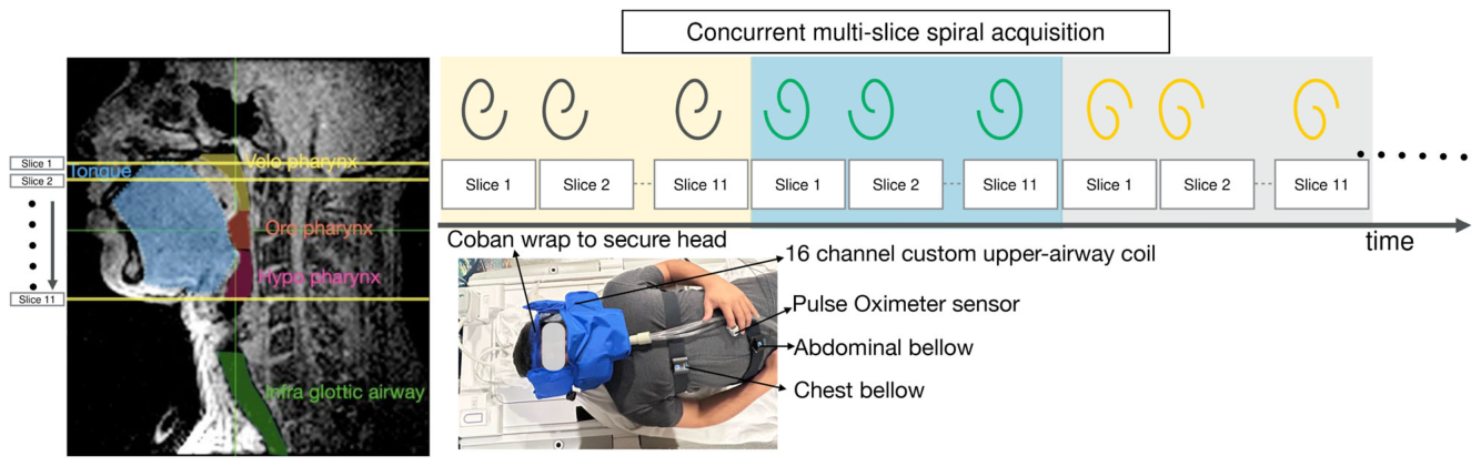
Figure 2. FIGURE 2 | Prospective acquisition setup and concurrent multi-slice spiral sampling scheme. (Left) Sagittal localizer showing the 11 axial slice locations spanning the velopharynx, oropharynx, hypopharynx. (Right, top) Concurrent multi-slice spiral acquisition: For each spiral arm angle, all 11 slices are acquired sequentially before incrementing to the next arm angle, yielding synchronized dynamic coverage across the full upper airway. (Right, bottom) Subject setup showing the 16-channel custom upper-airway receiver coil, Coban head wrap for head motion stabilization, pulse oximeter, and respiratory monitoring via abdominal and chest bellows.解説: 前向き収集のセットアップと同時多スライススパイラルサンプリングスキームを示しています。(左)11枚の軸方向スライスが口蓋咽頭から下咽頭までをカバーする位置を示しています。(右上)同時多スライススパイラル収集の概要で、各スパイラルアーム角度ごとに11枚の全スライスが順次収集される様子です。(右下)対象者のセットアップとして、カスタム16チャンネル上気道受信コイル、頭部安定化のためのコバンラップ、パルスオキシメーター、呼吸モニタリング用の腹部および胸部ベローが示されています。
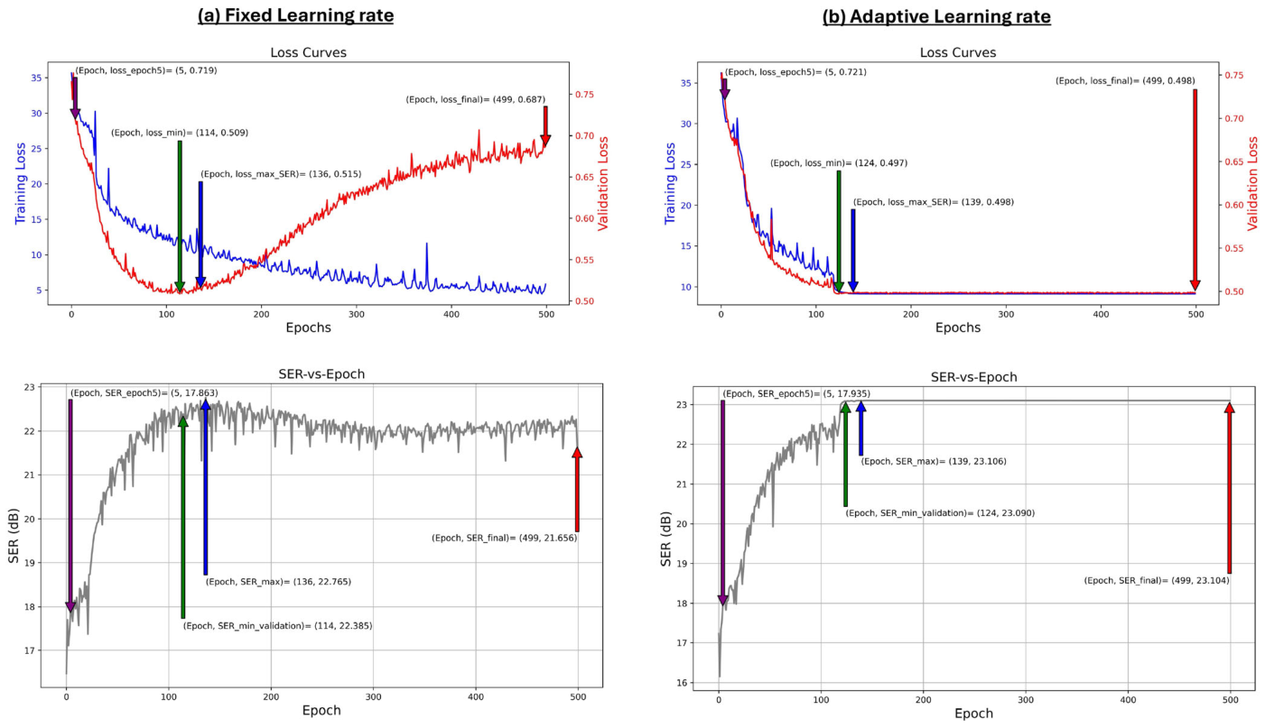
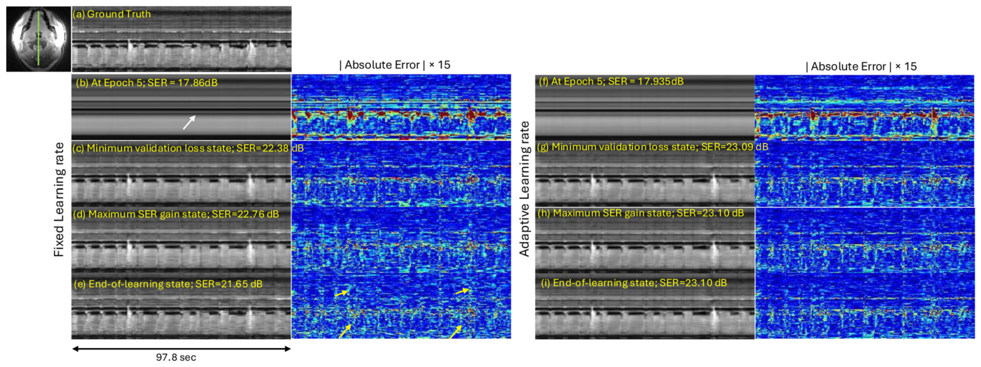
Figure 3. FIGURE 3 | Quantitative training dynamics under fixed and adaptive learning rate strategies. In each panel, the vertical arrows (from left to right) show epoch 5, minimum validation loss epoch, maximum SER-gain epoch state, final epoch state. (a) Fixed learning rate: Training and validation loss curves diverge after epoch 114, with SER peaking at 22.76 dB (epoch 136) before degrading to 21.65 dB at the final epoch. (b) Adaptive learning rate: Training and validation losses remain coupled throughout, with SER stable across the minimum-validation-loss (23.09 dB), maximum-SER-gain (23.10 dB), and final (23.10 dB) states.解説: 固定学習率戦略と適応学習率戦略における定量的訓練動態を比較しています。各パネルの垂直矢印は、エポック5、最小検証損失エポック、最大SERゲインエポック、最終エポックの状態を示します。(a)固定学習率では、訓練と検証損失曲線がエポック114以降で乖離し、SERは最終エポックで劣化します。(b)適応学習率では、訓練と検証損失は常に連動し、SERも安定しています。
Figure 4. FIGURE 4 | Qualitative reconstruction comparison at three learning states for fixed (left) and adaptive (right) learning rate strategies. Rows show mid-temporal cuts and corresponding 15× absolute error maps at (b, f) epoch 5, (c, g) minimum validation loss, (d, h) maximum SER gain, and (e, i) end of learning, against the ground truth reference (a). At epoch 5, the temporal information is not fully resolved (white arrow in (b)). Under the fixed learning rate, prolonged training introduces progressive noisy and blocky temporal artifacts (yellow arrows). The adaptive strategy maintains consistent reconstruction quality across all learning states.解説: 固定(左)と適応(右)学習率戦略における3つの学習状態での再構成の定性比較を示しています。各行は、(b, f)エポック5、(c, g)最小検証損失、(d, h)最大SERゲイン、(e, i)学習終了時の中間時間断面と、それに対応する15倍の絶対誤差マップを、グランドトゥルース(a)と比較して示しています。固定学習率では、訓練が長引くにつれてノイズやブロック状の時間的アーチファクト(黄色の矢印)が蓄積しますが、適応学習率戦略ではすべての学習状態を通じて一貫した再構成品質が維持されています。
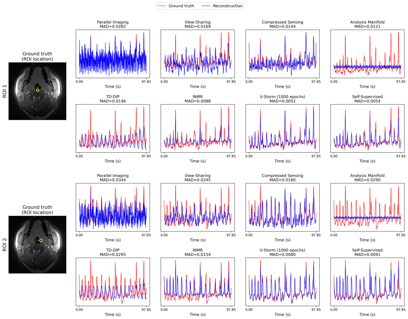
Figure 5. FIGURE 5 | Region-of-interest (ROI) time–intensity analysis on the retrospective dataset with ground truth. Two ROIs (yellow circles, left) were selected to capture regions of anterior–posterior and lateral dominant airway motion, respectively. Pixel time–intensity profiles (blue) are overlaid on the ground-truth profile (red) for each of the eight reconstruction methods: Parallel imaging, view sharing, compressed sensing, analysis manifold, TD-DIP, INMR, V-SToRM (1000 epochs), and the proposed self-supervised method. Mean absolute difference (MAD) between each reconstruction and the ground truth is annotated above each panel. The time axis spans the full retrospective acquisition (0–97.85 s). The proposed method and V-SToRM achieved the lowest MAD in both ROIs, with only a marginal difference between them (ROI 1: 0.0054 vs. 0.0052; ROI 2: 0.0091 vs. 0.0080). The remaining methods each showed a distinct failure mode: Parallel imaging exhibited pronounced noise amplification; view sharing, compressed sensing, and analysis manifold under-represented the rapid collapse peaks; TD-DIP similarly failed to capture rapid peaks while also showing residual blurring; and INMR, despite capturing rapid peaks in ROI 2, under-performed in ROI 1.解説: レトロスペクティブデータセットにおけるROI(関心領域)時間強度分析とグランドトゥルースの比較を示しています。(左の黄色い円)前後の動きが優勢な領域(ROI 1)と横方向の動きが優勢な領域(ROI 2)の2つのROIが選択されています。各パネルには、並列イメージング、ビューシェアリング、圧縮センシング、分析多様体、TD-DIP、INMR、V-SToRM(1000エポック)、提案された自己教師あり手法の8つの再構成方法について、ピクセル時間強度プロファイル(青色)がグランドトゥルースプロファイル(赤色)に重ねて表示されています。各再構成とグランドトゥルースとの間の平均絶対差(MAD)が各パネルの上に示されています。提案手法とV-SToRMが両方のROIで最も低いMADを達成しました。
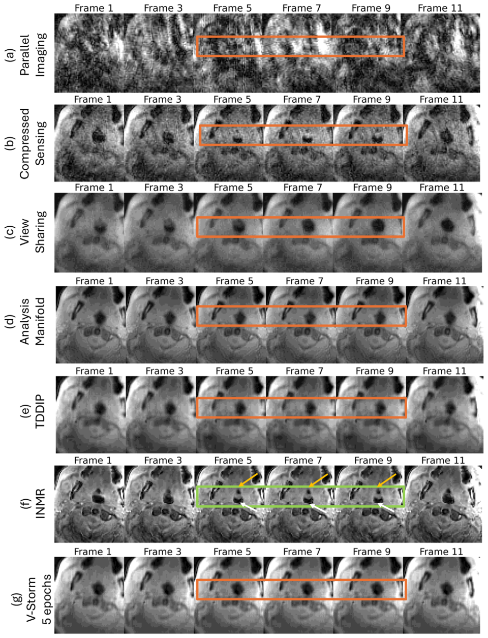
Figure 6. FIGURE 6 | Prospective reconstruction comparison across frames from a velopharyngeal slice of an OSA patient during natural sleep. (a) parallel imaging, (b) compressed sensing, (c) view sharing, (d) analysis manifold, (e) TD-DIP, (f) INMR, (g) V-SToRM (5 epochs), (h) V-SToRM (150 epochs), (i) V-SToRM (1000 epochs), (j) self-supervised reconstruction. Green boxes (frames 5–9) highlight the collapse interval where two anatomically distinct airway structures undergo dynamic narrowing: The retropalatal space posterior to the velum (white arrow) and the foramen cecum region anterior to the velum but behind the tongue (yellow arrow). Orange boxes denote regions affected by noise, spatially blurring, or temporal inconsistency. V-SToRM (150 epochs) improves temporal fidelity over V-SToRM (5 epochs), though has ambiguity in resolving the two airway structures in frames 7,9. V-SToRM (1000 epochs), INMR, and the proposed self-supervised method consistently resolve both structures across frames, though V-SToRM (1000 epochs) shows pronounced noise.解説: 自然睡眠中のOSA対象者の口蓋咽頭スライスからのフレームにわたる、前向き再構成の比較を示しています。(a)並列イメージング、(b)圧縮センシング、(c)ビューシェアリング、(d)分析多様体、(e)TD-DIP、(f)INMR、(g)V-SToRM (5エポック)、(h)V-SToRM (150エポック)、(i)V-SToRM (1000エポック)、(j)自己教師あり再構成です。緑色のボックス(フレーム5~9)は、口蓋帆後部のレトロパラタルスペース(白い矢印)と舌根部の盲孔領域(黄色の矢印)という2つの解剖学的に異なる気道構造が動的に狭窄する閉塞期間を強調しています。オレンジ色のボックスは、ノイズ、空間的ぼやけ、または時間的一貫性の欠如によって影響を受ける領域を示しています。V-SToRM(150エポック)はV-SToRM(5エポック)よりも時間的忠実度が向上していますが、フレーム7、9では2つの気道構造の解像に曖昧さがあります。V-SToRM(1000エポック)、INMR、および提案された自己教師あり手法は、フレーム全体で両方の構造を一貫して解像していますが、V-SToRM(1000エポック)では顕著なノイズが見られます。
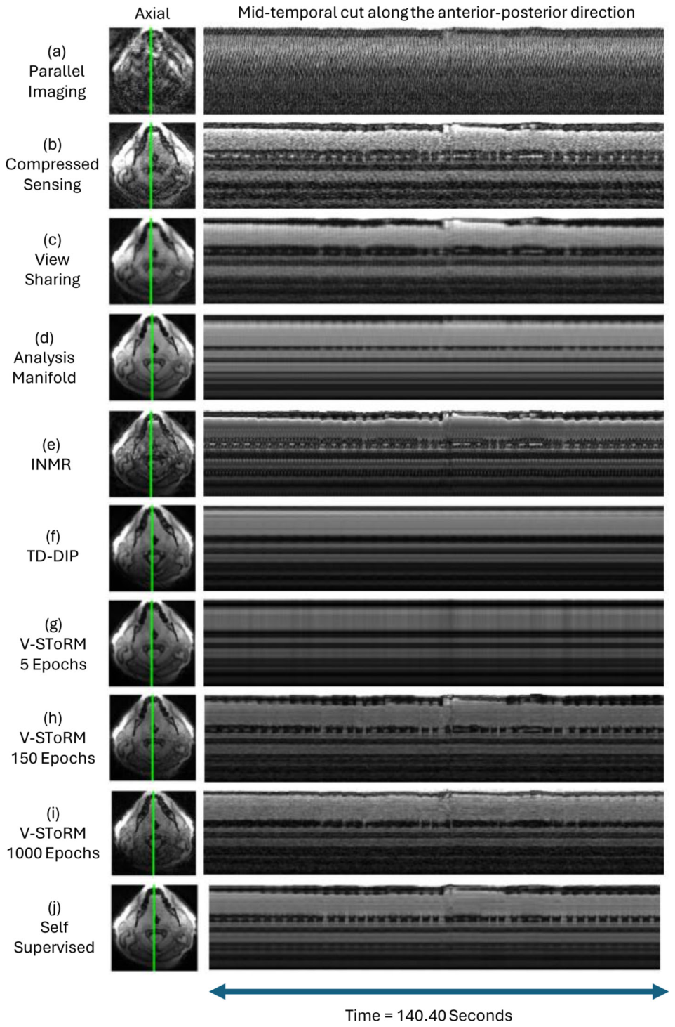
Figure 7. FIGURE 7 | Prospective reconstruction comparison from a velo-pharyngeal slice of a second OSA patient during natural sleep. The left column shows the axial profile with the green line indicating the mid-temporal cut location; the right column shows the corresponding spatiotemporal profile along the anterior–posterior direction for (a) parallel imaging, (b) compressed sensing, (c) view sharing, (d) analysis manifold, (e) INMR, (f) TD-DIP, (g) V-SToRM at 5 epochs, (h) V-SToRM at 150 epochs, (i) V-SToRM at 1000 epochs and (h) self-supervised reconstruction. Analysis manifold and TD-DIP over-smooth the temporal profile with loss of collapse dynamics; INMR shows prominent high-frequency noise throughout. V-SToRM at 5 epochs show loss of temporal detail. V-SToRM at 1000 epochs shows noisier profile characteristic of prolonged training. On this patient, V-SToRM at 150 epochs and the proposed method preserves reliable air–tissue boundaries and smooth, continuous temporal evolution. Representative comparisons on more OSA patient datasets are provided in the Figure S8.解説: 自然睡眠中の別のOSA対象者の口蓋咽頭スライスからの前向き再構成の比較を示しています。左の列は軸方向プロファイルで、緑色の線は中間時間断面の位置を示しています。右の列は、(a)並列イメージング、(b)圧縮センシング、(c)ビューシェアリング、(d)分析多様体、(e)INMR、(f)TD-DIP、(g)V-SToRM 5エポック、(h)V-SToRM 150エポック、(i)V-SToRM 1000エポック、(j)自己教師あり再構成に対応する前後方向の時間空間プロファイルを示しています。分析多様体とTD-DIPは時間プロファイルを過度に滑らかにし、閉塞動態が失われています。INMRは全体的に高い周波数ノイズを示します。V-SToRM 5エポックは時間的詳細が失われ、V-SToRM 1000エポックは訓練の長期化に特徴的なノイズの多いプロファイルを示します。この対象者では、V-SToRM 150エポックと提案手法が、信頼性の高い空気と組織の境界と滑らかで連続的な時間的変化を保持しています。
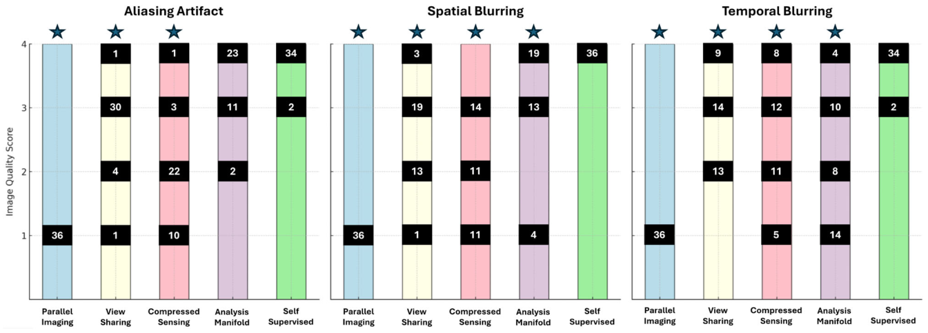
Figure 8. FIGURE 8 | Expert image quality scores across 12 datasets (8 OSA patients during natural sleep and 4 healthy volunteers performing the Müller maneuver) for five reconstruction methods, evaluated independently by three expert raters—an otolaryngologist, a radiologist, and a sleep medicine physician—yielding 36 total ratings per method. Stacked bars show the distribution of scores (1 = unacceptable, 4 = excellent) for aliasing artifacts, spatial blurring, and temporal blurring. Stars denote the highest-scoring method in each category. Kruskal–Wallis tests confirmed significant differences among methods in all categories (𝑝< 0.001); pairwise Mann–Whitney U tests confirmed that the self-supervised method significantly outperformed all competing approaches (𝑝< 0.05).解説: 12のデータセット(自然睡眠中のOSA対象者8名とミュラー手技を行う健康なボランティア4名)における5つの再構成方法に対する専門家による画像品質スコアを示しています。これは、耳鼻咽喉科専門家、放射線科専門家、睡眠医学専門家の3名の専門家によって独立して評価され、各方法につき合計36の評価が得られました。積み上げ棒グラフは、エイリアシングアーチファクト、空間的ぼやけ、時間的ぼやけの3つのカテゴリーにおけるスコア(1=許容できない、4=優れている)の分布を示しています。星印は各カテゴリーで最高スコアを獲得した方法を示します。自己教師あり再構成は、すべてのカテゴリーで競合するすべての方法よりも有意に優れていると評価されました。
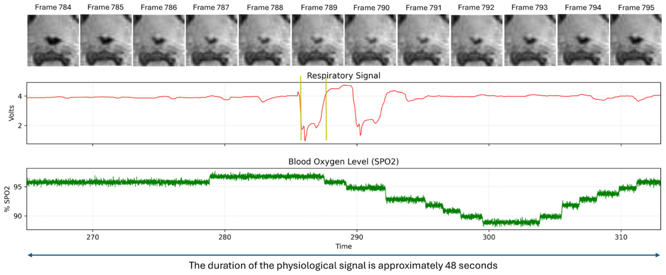
Figure 9. FIGURE 9 | Self-supervised reconstructed frames from the velo-pharynx of an OSA patient during natural sleep, alongside concurrent respiratory effort (red) and SpO2 (green) over a 48-s window. Vertical yellow lines mark the ≈2-s collapse interval, during which respiratory effort drops and then surges followed by SpO2 desaturation from 96% to < 89%. The delay between collapse and SpO2 nadir reflects buffering by the lung oxygen reservoir. Reconstructed frames show progressive velo-pharyngeal narrowing in temporal correspondence with the physiological signals.解説: 自然睡眠中のOSA対象者の口蓋咽頭から自己教師あり再構成されたフレームと、同期した呼吸努力(赤色)およびSpO₂(緑色)の48秒間のデータを並べて示しています。垂直の黄色の線は、約2秒間の閉塞期間を示しており、この間、呼吸努力は低下した後、急増しています。これに続いてSpO₂が96%から89%未満に低下しており、気道狭窄とSpO₂の最低値との時間的遅延は肺の酸素貯蔵による緩衝を反映しています。再構成されたフレームは、生理学的信号と時間的に対応する口蓋咽頭の進行性の狭窄を示しています。
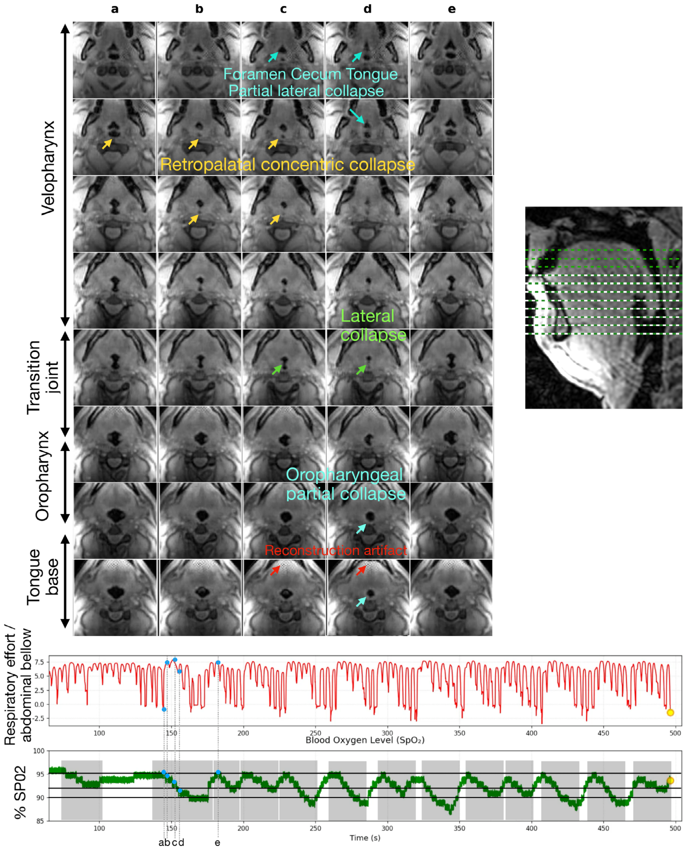
Figure 10. FIGURE 10 | Multi-site upper-airway collapse dynamics in an OSA patient with a large velum during natural sleep. (Bottom) Respiratory effort (red) and SpO2 (green) over a single ≈8-min run; gray shading marks 13 identified collapse events (≥3% SpO2 drop). Dashed lines correspond to timepoints a–e. (Top) 8 of the 11 contiguous axial slices at five timepoints (a–e) spanning a collapse and recovery sequence across the velo-pharynx, transition joint, oropharynx, and tongue base. Colored arrows indicate: Complete concentric retro-palatal collapse (yellow), lateral collapse at the velo-pharyngeal to oro-pharyngeal transition (green), partial lateral collapse at the foramen cecum of the tongue (cyan), and oro-pharyngeal partial lateral collapse (blue). Red arrows indicate localized reconstruction artifacts; further examples in Figure S6. (Right) Sagittal localizer showing 11 contiguous slice locations. The yellow circle indicates the end of the 8-min run.解説: 自然睡眠中の大口蓋帆を有するOSA対象者における、多部位上気道閉塞動態を示しています。(下) 約8分間の単一スキャンにおける呼吸努力(赤色)とSpO₂(緑色)のデータです。灰色の網掛けは、特定された13回の閉塞イベント(3%以上のSpO₂低下)を示しています。点線は時間点a~eに対応します。(上) 口蓋咽頭、移行部、中咽頭、舌根部にわたる閉塞と回復のシーケンスを示す5つの時間点(a~e)での、11枚の連続軸方向スライスのうち8枚を示しています。色付きの矢印は、完全な同心円状口蓋帆後部閉塞(黄色)、口蓋咽頭から中咽頭への移行部での側壁閉塞(緑色)、舌の盲孔部分での部分的な側壁閉塞(シアン)、中咽頭の部分的な側壁閉塞(青色)を示しています。赤色の矢印は局所的な再構成アーチファクトを示しています。(右) 11枚の連続スライス位置を示す矢状方向の局所化画像です。この図は、自然睡眠中の複雑な気道閉塞の動態を多レベルで詳細に可視化できることを示しています。

コメントを残す