どうも、Beyond the Pixelです。乳房MRIは、乳がんの検出と診断において重要な役割を果たしています。通常、この検査ではガドリニウム系造影剤(GBCA)を用いてダイナミック造影MRI(DCE-MRI)を実施します。これにより、組織の造影パターンが高解像度で捉えられ、病変の検出と特性評価が可能になります。しかし、GBCAの使用には、準備や実施に手間がかかることや、環境への影響が懸念されるといった課題があります。また、ガドリニウム曝露に関連する症状(SAGE)についても研究が進められており、造影剤を使用しないMRIアプローチへの関心が高まっています。
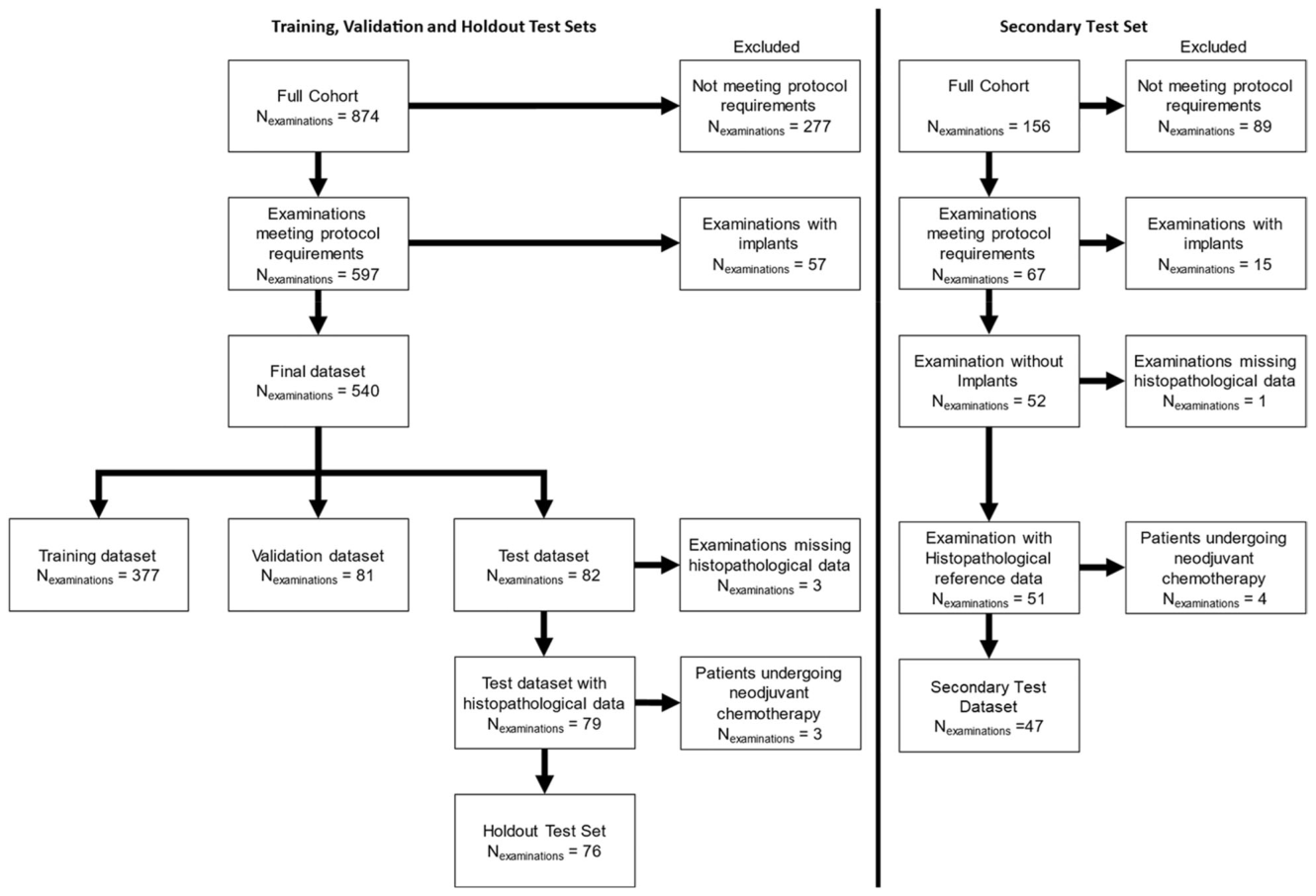
Figure 1. Fig. 1 The full cohort used for the creation of the training, validation and holdout sets consisted of patients undergoing breast MRI on a single Siemens Healthineers MAGNETOM Skyra Fit scanner. The full cohort for the creation of the secondary test set consisted of consecutive breast MRI examinations on a MAGNETOM VIDA scanner between July 2018 and January 2019. The protocol was defined as including a five-point DCE acquisition, a T1W acquisition, a T2W fat-saturated acquisition and a multi-b-value diffusion-weighted MRI (DWI) acquisition containing b-values of 50, 750, and 1500 s/mm2解説: 本図は、研究で使用された対象者コホートの選択プロセスを示しています。トレーニング、検証、ホールドアウトテストセットの作成に使用された全コホートと、セカンダリテストセットの作成に使用された全コホートが、除外基準を経て最終的なデータセットに分割される流れが示されています。
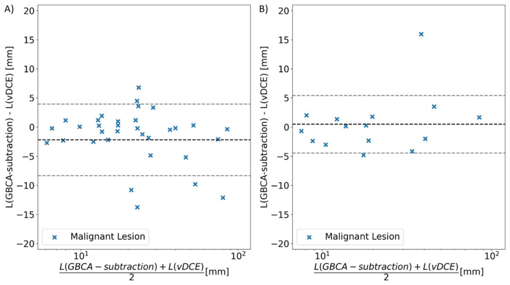
Figure 5. Fig. 5 Bland–Altman plot comparison of length of the longest axis (L) of malignant findings measured either on the GBCA-subtraction or on the vDCE series for holdout (A) and secondary (B) test set. The mean paired differences were −2.2 mm and 0.5 mm, corresponding to mean vDCE overestimation in the holdout test set of 2.2 mm and underestimation of 0.5 mm in the secondary test set. GBCA, gadolinium-based contrast agent; vDCE, virtual dynamic contrast-enhanced解説: 本図は、悪性病変の最長軸長(L)の測定値について、GBCA減算画像またはvDCEシリーズで測定した場合のBland–Altmanプロット比較を示しています。Aはホールドアウトテストセット、Bはセカンダリテストセットのデータです。平均的な病変サイズの差(DCEからvDCEを引いた値)は、ホールドアウトテストセットで-2.2mm、セカンダリテストセットで0.5mmでした。
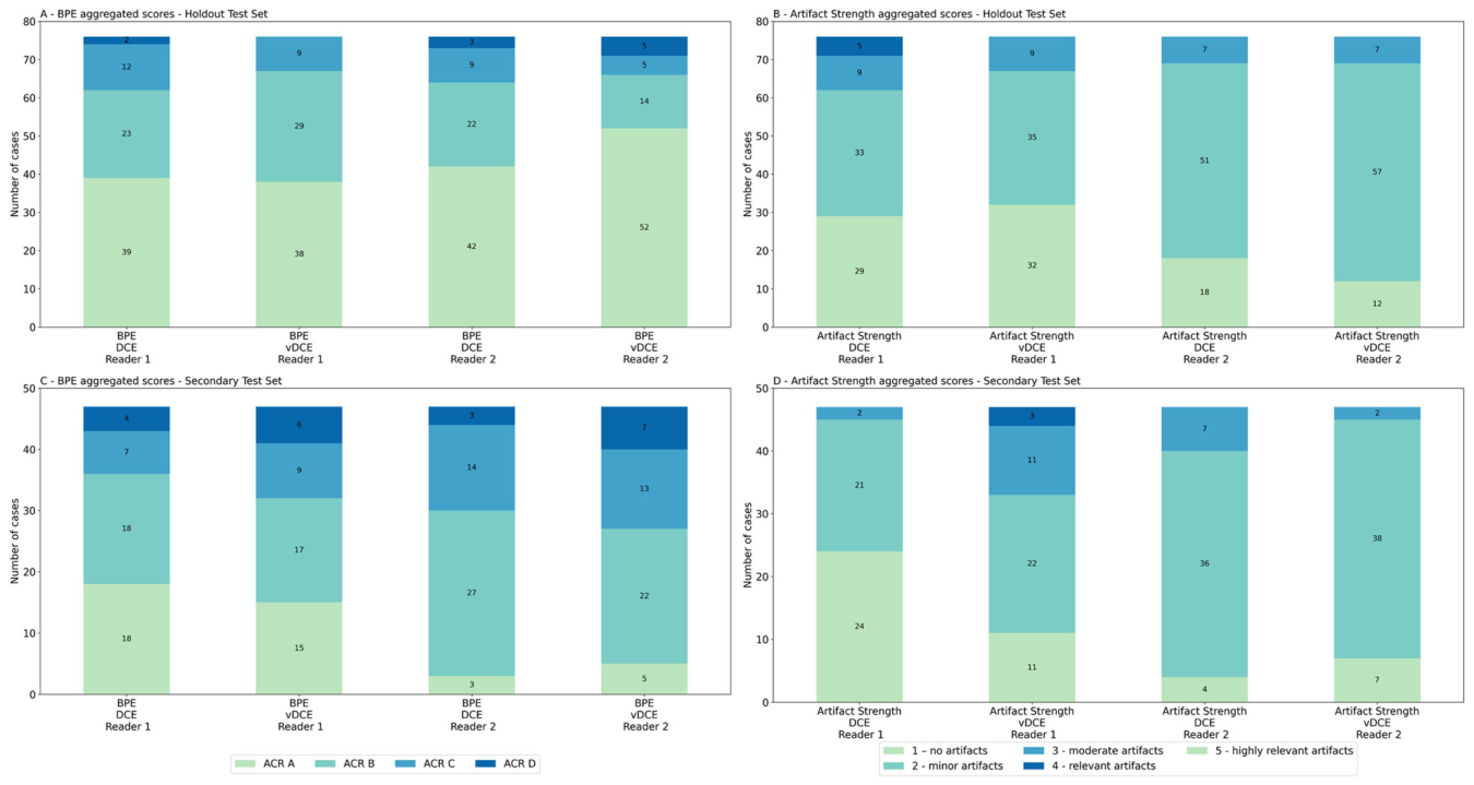
Figure 6. Fig. 6 Aggregated scores for BPE and Artefact strength for the holdout (A, B) and secondary (C, D) test datasets解説: 本図は、ホールドアウトテストセット(A, B)とセカンダリテストセット(C, D)において、2名の専門家が評価した背景乳腺実質濃染(BPE)とアーチファクト強度の集計スコアの分布を示しています。
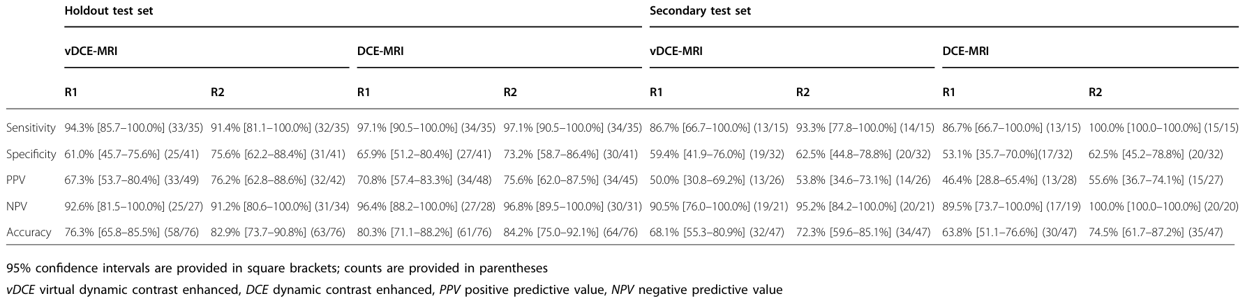
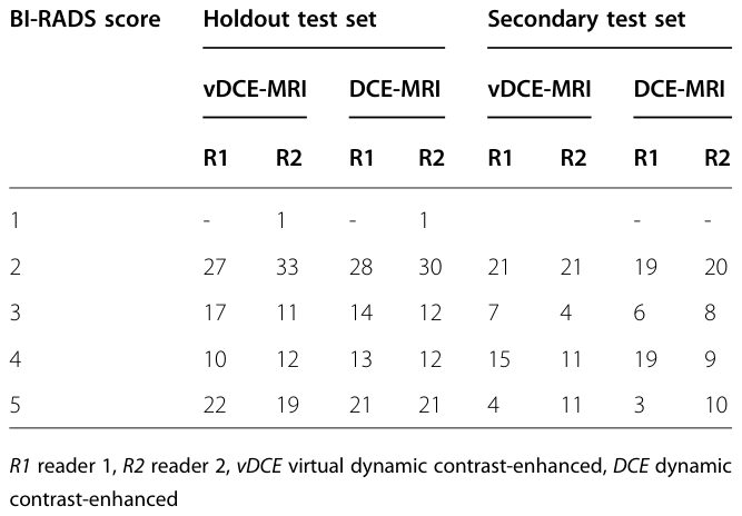
Table 4. Table 4 Aggregated BI-RADS scores in test sets解説: 本表は、ホールドアウトテストセットとセカンダリテストセットにおいて、2名の専門家(R1, R2)がvDCE-MRIとDCE-MRIで割り当てたBI-RADSスコアの集計値を示しています。各スコア(1から5)のケース数が記載されています。
また、従来のDCE画像とvDCE画像では、病変の描出に違いが見られるケースもありました。
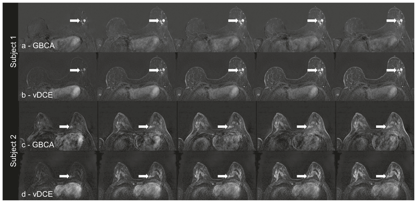
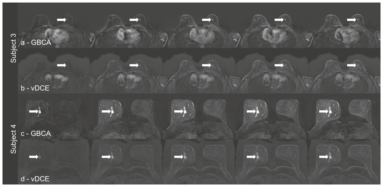
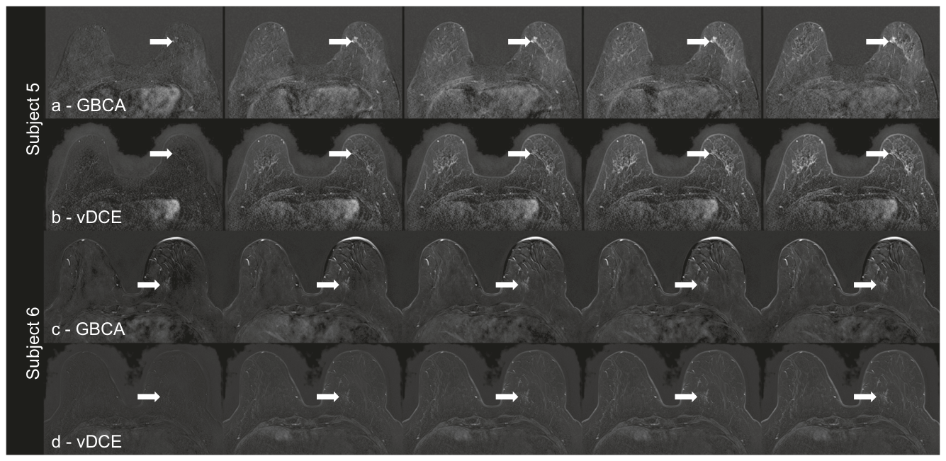
Figure 2. Fig. 2 GBCA-subtraction (a, c) and vDCE (b, d) series (b, d) for two subjects from the holdout test set. Subject 1 is a 71-year-old woman undergoing diagnostic breast MRI due to suspicion of multifocal breast cancer on X-ray mammography. The vDCE-subtraction and the GBCA-subtraction depict suspicious lesions spreading over an area of 24 mm in the left breast, as marked by the arrow. Biopsy confirmed an invasive breast carcinoma (NST). Subject 2 is a 49-year-old woman undergoing diagnostic breast MRI due to suspicion of multifocal disease on X-ray mammography. The vDCE subtraction and the GBCA subtraction depict increased background parenchymal enhancement, with the vDCE visualisation of the BPE appearing patchier and less granular than after GBCA administration. Despite the BPE, a suspicious lesion of 6x8mm in the left breast was identified(arrow) on GBCA and vDCE. Biopsy confirmed an invasive breast carcinoma (NST). FGT, fibroglandular tissue; GBCA, gadolinium-based contrast agent; vDCE, virtual dynamic contrast-enhanced解説: 本図は、ホールドアウトテストセットの2名の対象者における、GBCAによる減算画像(a, c)とvDCE画像(b, d)の比較を示しています。対象者1では、両画像で左乳房に24mmの領域に広がる疑わしい病変が示され、生検で浸潤性乳がんが確認されました。対象者2では、両画像で背景乳腺実質濃染の増加が描出されており、vDCEの方がパッチ状で粒状性が低いものの、左乳房に6x8mmの疑わしい病変が特定され、生検で浸潤性乳がんが確認されました。Figure 3. Fig. 3 GBCA-subtraction (a, c) and vDCE (b, d) series (b, d) for two subjects from the secondary test set. Subject 3: a 60-year-old woman undergoing diagnostic breast MRI. An 8×8 mm non-mass enhancement can be noted in both the DCE and vDCE series (white arrow). Histopathology confirmed invasive breast carcinoma (NST). Subject 4 is a 56-year-old woman undergoing diagnostic breast MRI due to suspicion of multifocal breast cancer. Both vDCE and GBCA subtraction images present multiple lesion clusters, with the largest lesion being 16 x 11 mm. Histopathology confirmed invasive breast carcinoma (NST) with ductal carcinoma in-situ (DCIS) components. FGT, fibroglandular tissue; GBCA, gadolinium-based contrast agent; vDCE, virtual dynamic contrast-enhanced解説: 本図は、セカンダリテストセットの2名の対象者における、GBCAによる減算画像(a, c)とvDCE画像(b, d)の比較を示しています。対象者3では、DCEとvDCEの両方で8x8mmの非腫瘤性濃染が確認され、生検で浸潤性乳がんが確認されました。対象者4では、両画像で複数の病変クラスターが提示され、最大の病変は16x11mmであり、生検で浸潤性乳がんが確認されました。Figure 4. Fig. 4 GBCA-subtraction (a, c) and vDCE series (b, d) for one subject from the holdout test set (Subject 5) and one subject from the secondary test set (Subject 6). Subject 5 is a 61-year-old woman undergoing diagnostic breast MRI. Whilst the GBCA-subtraction depicts a suspicious non-mass enhancement of 8 × 9mm in the left breast (arrow), the respective region was rated not suspicious for both readers in the vDCE-subtraction, as the lesion was interpreted as part of the adjacent BPE. Biopsy confirmed an invasive breast carcinoma (NST). Subject 6 was a 60-year-old woman undergoing diagnostic breast MRI. The suspicious non-mass enhancement (28 × 17mm) was correctly identified in the GBCA-subtraction series by both readers. However, in the vDCE series, only one reader identified the non-mass enhancement correctly. Histopathology confirmed invasive breast carcinoma (NST). FGT, fibroglandular tissue; GBCA, gadolinium-based contrast agent; vDCE, virtual dynamic contrast-enhanced解説: 本図は、ホールドアウトテストセットの対象者5とセカンダリテストセットの対象者6における、GBCAによる減算画像(a, c)とvDCE画像(b, d)の比較を示しています。対象者5では、GBCA減算画像で8x9mmの疑わしい非腫瘤性濃染が示されたのに対し、vDCE減算画像では両専門家によってこの病変は疑わしいとは評価されませんでした。生検で浸潤性乳がんが確認されました。対象者6では、GBCA減算画像で28x17mmの疑わしい非腫瘤性濃染が両専門家によって正しく特定されましたが、vDCEシリーズでは1名の専門家のみが正しく特定しました。生検で浸潤性乳がんが確認されました。


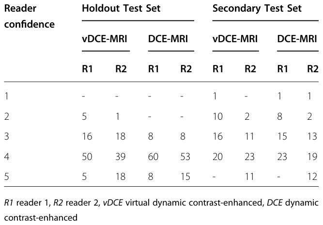
コメントを残す