どうも、Beyond the Pixelです。慢性閉塞性肺疾患(COPD)は世界中で4億人以上が罹患し、主要な死因の一つとなっています。気道の粘液病態は気流閉塞に寄与し、治療標的となる可能性が注目されています。COPDでは、粘液線毛機能不全により粘液が濃縮され、粘稠性が増し、排出が阻害されます。これは気道を閉塞する粘液栓の形成につながり、気流閉塞や増悪、死亡率の上昇といった臨床的転帰不良に関連しています。これまでの研究では、胸部CTが粘液栓を特定するための実用的なアプローチとして浮上しており、COPDコホートの約40〜60%で粘液栓が報告されています。CTで「サイレント」な粘液栓がある場合でも、機能障害や増悪と関連していることが示されています。近年では、このような粘液栓の検出を自動化し、大規模に適用するためにAIベースの自動検出が開発されています。自動的な粘液栓検出は、臨床診療や多施設共同研究において、気道閉塞の標準化された評価を可能にする可能性があります。しかし、診断性能に関する独立した検証は限られており、自動検出が臨床的に関連する気道疾患をどの程度反映しているかについては、さらなる明確化が必要です。この研究では、胸部CTにおける自動粘液栓検出アルゴリズムを、専門家による評価と比較し、AIが専門家の検出能力を向上させるかどうかを評価し、アルゴリズム由来の気道閉塞の負担と臨床的および機能的重症度との関連を調べました。
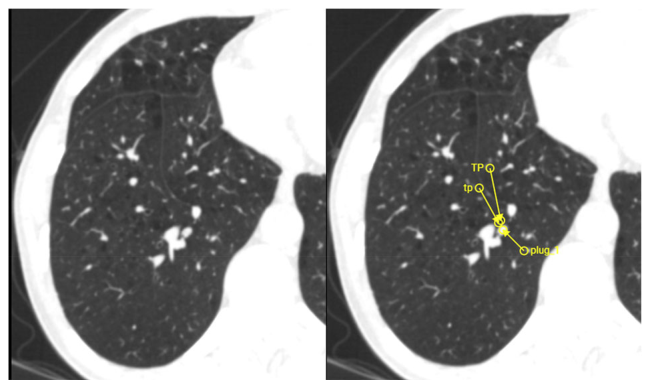
Figure 1. Fig. 1 Representative example of mucus plug annotation and AI- assisted candidate review. The unannotated axial CT image (left) shows complete obstruction of a posterior basal segmental bronchus by a mucus plug in the right lower lobe. The annotated image (right) demonstrates lesion-level localization using arrow annotations, with the arrowheads placed at the site of obstruction. Human reader annotations (mp, TP) and the AI-generated candidate marker (plug_1) localizes the same mucus plug, which was adjudicated as a true- positive finding. In the AI-assisted session, AI candidates were displayed as in-slice markers in the same viewer, allowing readers to review the corresponding axial image and adjacent slices before accepting or rejecting each candidate解説: この図は、胸部CT画像における粘液栓の検出例を示しています。左側は注釈のないCT画像で、右側は専門家とAI(plug_1)の両方によって同じ粘液栓が検出され、注釈が付けられた画像です。これは右下葉の気管支腔が粘液栓により完全に閉塞していることを示しています。
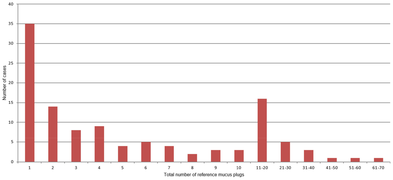
Figure 2. Fig. 2 Distribution of mucus plugs per case (n = 114). Bar chart showing the distribution of the total number of adjudicated reference mucus plugs identified per case. The x-axis indicates the total number of adjudicated reference mucus plugs, with cases containing more than ten mucus plugs grouped into predefined ranges (11–20, 21–30, 31–40, 41–50, 51–60, and 61–70). Most cases had relatively few mucus plugs, while a smaller subset exhibited a high mucus plug burden解説: この棒グラフは、対象者(n=114)ごとに判定された参照粘液栓の総数の分布を示しています。ほとんどの対象者では比較的少数の粘液栓が見られましたが、一部の対象者では多数の粘液栓が存在することがわかります。
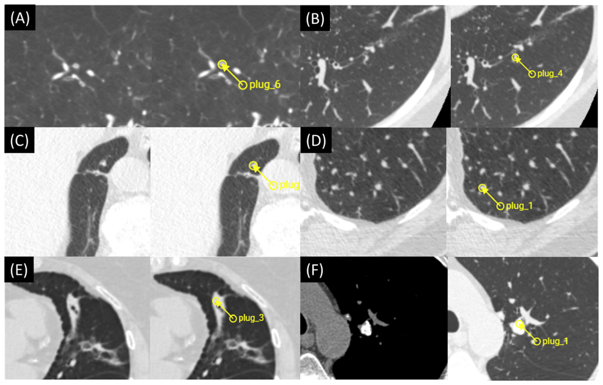
Figure 3. Fig. 3 Representative examples of AI detections not meeting the reference definition of complete mucus plugs. A Concentric bronchial wall thickening with a preserved pin-point lumen. B Partial mucus plug causes luminal obstruction in the lateral part of the bronchus, with a thin-walled patent lumen medially. C Mucus plug detected in the right lower lobe at the azygoesophageal recess within 2 cm of the pleura. D Peripheral pulmonary artery misclassified as a mucus plug. E Abrupt termination of a bronchiolectatic lumen due to focal consolidation. F Calcified endobronchial hamartoma misclassified as a mucus plug解説: この図は、完全な粘液栓の参照定義を満たさなかったAI検出の代表的な例を示しています。(A) 狭い内腔が保たれた同心円状の気管支壁肥厚。(B) 気管支の一部が部分的に閉塞した粘液栓。(C) 胸膜から2cm以内の右下葉で検出された粘液栓。(D) 粘液栓と誤分類された末梢肺動脈。(E) 巣状の浸潤影による細気管支拡張した内腔の突然の終末。(F) 粘液栓と誤分類された石灰化を伴う気管支内過誤腫。
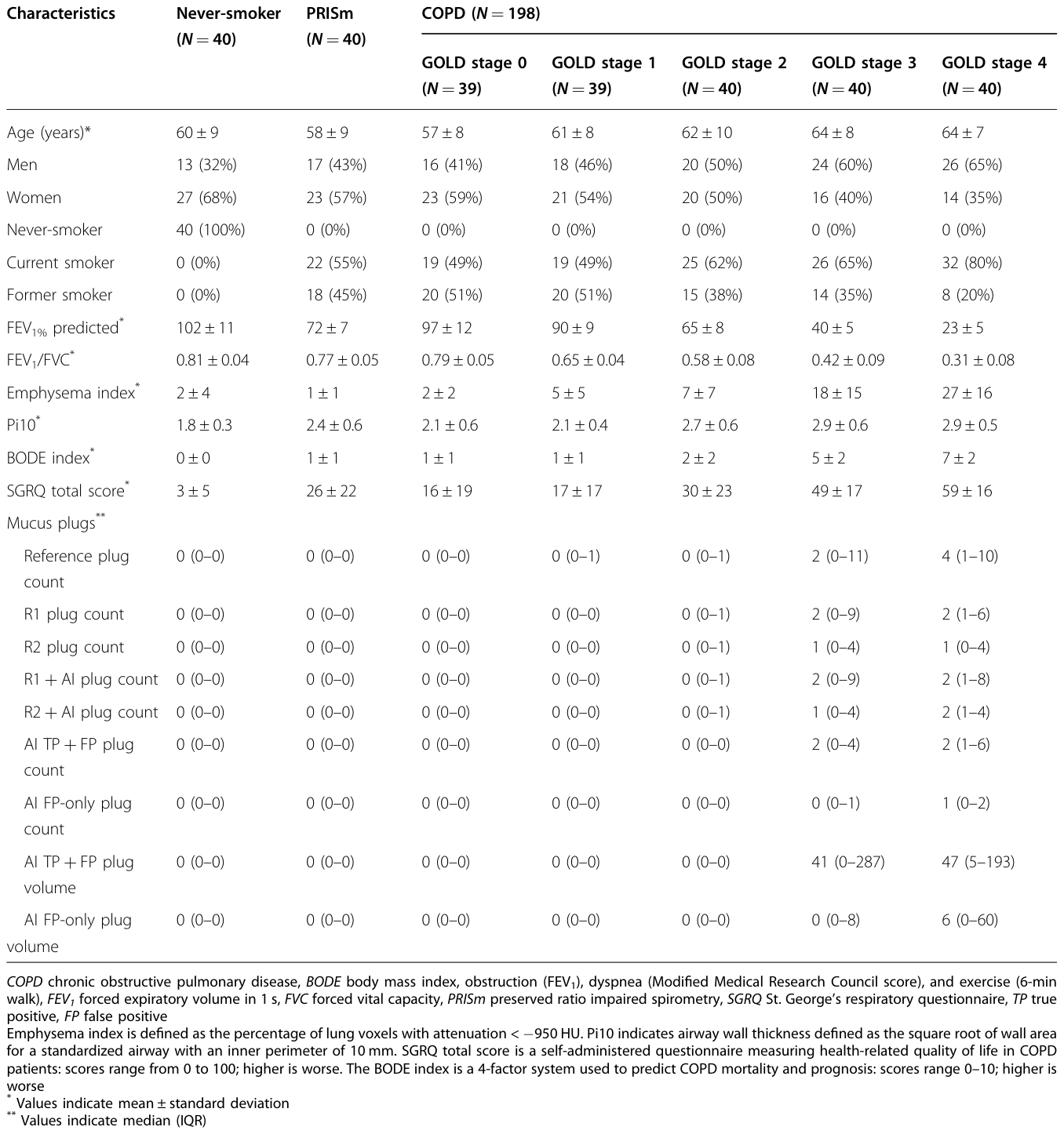
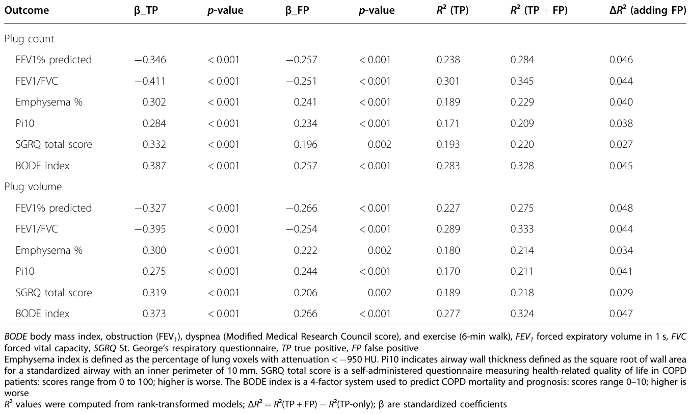
Table 4. Table 4 Associations of AI plug count and volume with outcomes in regression models including TP-only burden and additional non-reference-positive AI detections解説: この表は、真陽性(TP)のみの負担と「参照標準にない陽性」のAI検出を含む回帰モデルにおける、AI粘液栓カウントおよびボリュームと臨床的アウトカムとの関連性を示しています。FEV1%予測値、FEV1/FVC、肺気腫%、Pi10、SGRQ合計スコア、BODE指数といったアウトカムに対するTPおよびFPのβ値とp値、そしてFPの追加がモデル適合度(R²)に与える影響(ΔR²)が記載されています。


コメントを残す