どうも、Beyond the Pixelです。肝硬変に起因する門脈圧亢進症は、重篤な合併症を引き起こし、対象者の罹患率や死亡率に大きく影響します。この状態はしばしば心拍出量の増加、全身血管抵抗の低下、動脈性低血圧を特徴とする過動循環を伴います。経頸静脈的肝内門脈大循環シャント術(TIPS)は、門脈圧亢進症の治療として確立された選択肢です。TIPSは門脈と全身静脈系との間に低抵抗のバイパスを形成し、門脈循環を効果的に減圧します。これにより、出血性食道静脈瘤や難治性腹水などの合併症を改善する効果が期待されます。しかし、門脈減圧は脾臓領域の血液量を急性的に全身循環に再分配し、心臓への前負荷を増加させ、右心房圧や肺動脈圧、心臓の拡張を招くことが知られています。この容量過負荷は、心臓の過動状態をさらに悪化させる可能性があります。これまでの研究では、TIPS留置後の心臓への影響は主に短期的または中期的な機能的変化に焦点が当てられており、長期的な心臓の形態、機能、心筋組織特性の変化については十分に解明されていませんでした。心臓MRIは、心室容積や収縮機能を包括的かつ再現性高く評価できるだけでなく、T1マッピングやT2マッピングといった高度な心筋組織特性評価を可能にし、びまん性心筋浮腫や線維化の非侵襲的検出に役立ちます。今回の研究では、肝硬変による門脈圧亢進症の対象者におけるTIPS留置後の心臓MRIによる機能的および組織的パラメータの長期的な変化を評価することを目的としました。
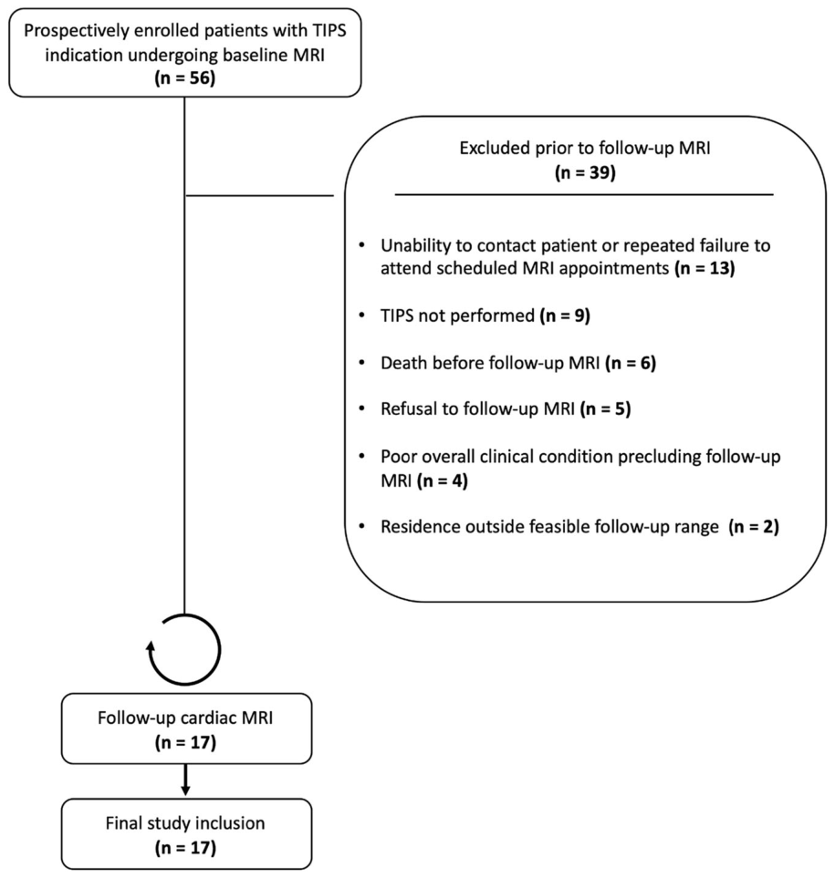
Figure 1. Fig. 1 Study flowchart. Seventeen patients underwent baseline MRI prior to transjugular intrahepatic portosystemic shunt (TIPS) insertion and follow- up MRI and were included in the final analysis解説: TIPS(経頸静脈的肝内門脈大循環シャント術)の適応があり、ベースラインMRIを受けた登録対象者の研究フローチャートを示します。フォローアップMRI前に39名が除外され、最終的に17名の対象者が研究に含まれました。
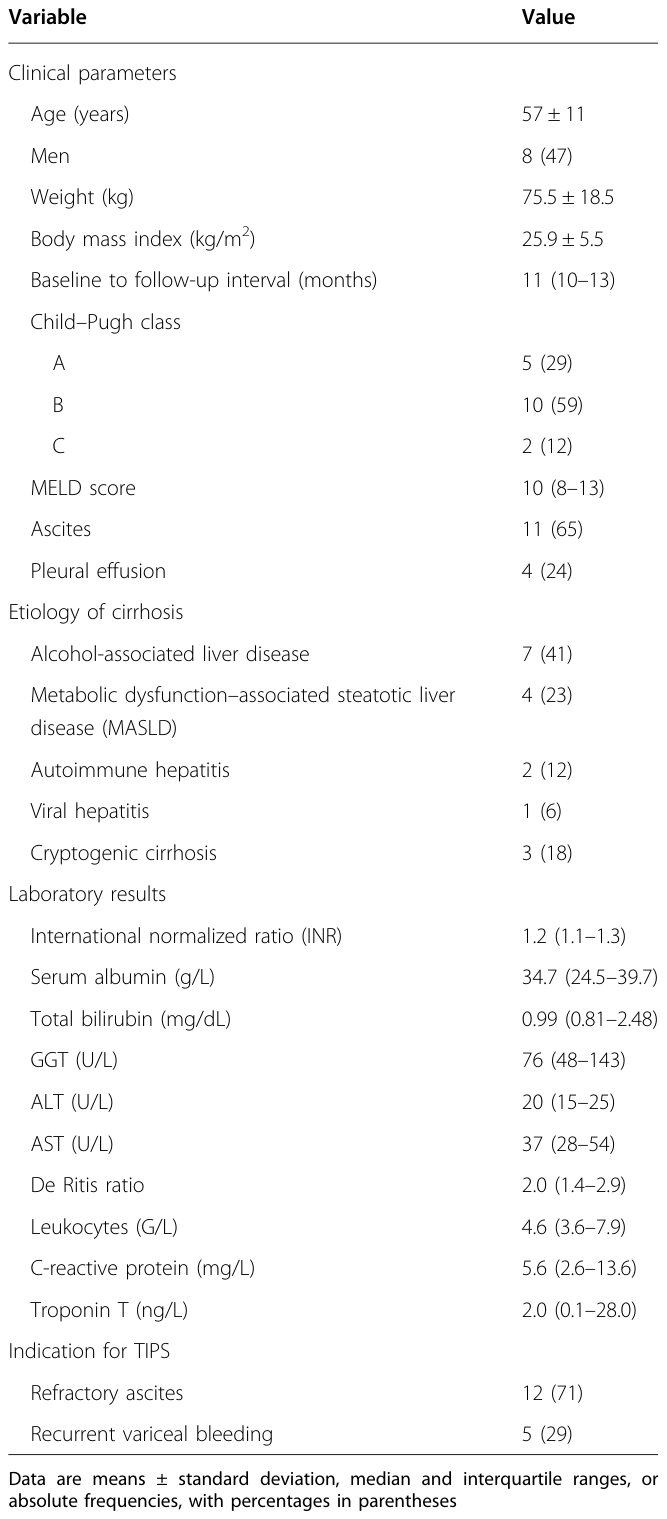
Table 1. Table 1 Clinical characteristics of the study cohort at baseline CMR (n = 17)解説: 研究コホート(17名)のベースライン心臓MRI時の臨床特性を示しています。対象者の年齢、性別、BMI、Child–Pugh分類、MELDスコア、腹水・胸水の有無、肝硬変の原因、およびTIPSの適応(難治性腹水または再発性静脈瘤出血)がまとめられています。また、INR、血清アルブミン、総ビリルビンなどの検査値も含まれています。
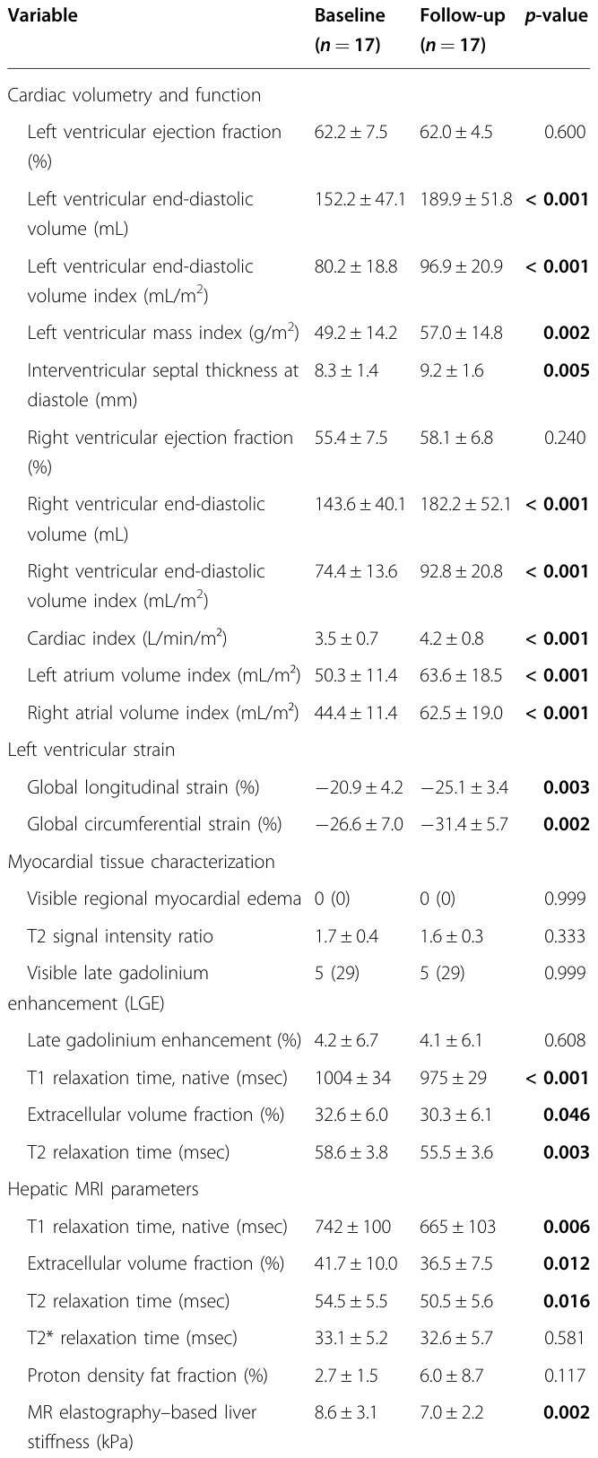
Table 3. Table 3 MRI results at baseline and on follow-up after transjugular intrahepatic portosystemic shunt (TIPS) procedure解説: TIPS処置後のベースラインとフォローアップにおけるMRI結果を示しています。心臓の容量と機能(左心室駆出率、拡張末期容積、心筋量指数、心臓指数など)、左心室ストレイン(グローバル縦方向ストレイン、グローバル円周方向ストレイン)、心筋組織特性(T1/T2緩和時間、ECV)、肝臓MRIパラメータ、脾臓MRIパラメータがまとめられています。
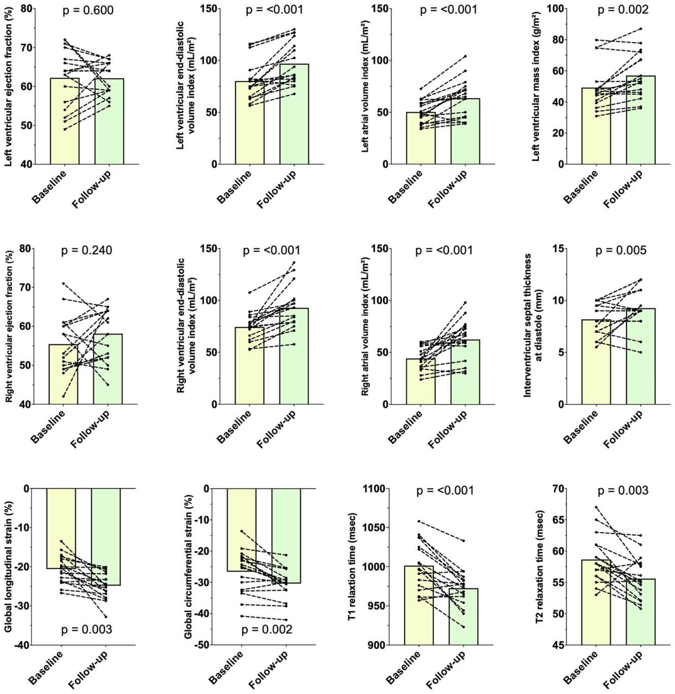
これらの心臓パラメータの変化は、
Figure 2. Fig. 2 Bar graphs show changes in cardiac parameters between baseline and follow-up MRI. Intra-individual values are plotted between both examinations. For intra-individual comparison, paired t-test or Wilcoxon signed-rank test was used解説: ベースラインMRIとフォローアップMRIにおける心臓パラメータの変化を棒グラフで示しています。各対象者の値が両検査間でプロットされており、心臓容積の増加、心筋ストレインの改善、T1およびT2緩和時間の減少が視覚的に示されています。
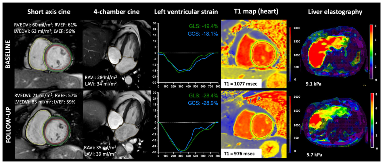
Figure 3. Fig. 3 Clinical example of a 59-year-old woman with alcohol-associated liver cirrhosis (Child–Pugh class B; Model for End-stage Liver Disease (MELD) score 8) who underwent baseline MRI 5 days prior to TIPS insertion and follow-up MRI 10 months thereafter. Short-axis and 4-chamber cine imaging demonstrate a longitudinal increase in right (RAVi) and left atrial volume index (LAVi), as well as right (RVEDVi) and left ventricular end-diastolic volume index (LVEDVi), reflecting sustained cardiac volume loading at follow-up. Despite these volumetric changes, right (RVEF) and left ventricular ejection fractions (LVEF) remained largely preserved. However, left ventricular strain analysis reveals marked improvement in both global longitudinal strain (GLS) and global circumferential strain (GCS), indicating recovery of pre-existing subclinical systolic dysfunction. Consistent with functional improvement, myocardial T1 mapping demonstrates normalization of quantitative tissue characterization parameters from elevated baseline values to follow-up (center-specific cutoff: T1 ≥1000 msec), suggesting resolution of diffuse myocardial edema. MR liver elastography at comparable anatomical slices shows a concomitant reduction in liver stiffness, supporting a systemic effect of portal decompression on tissue hydration and解説: アルコール性肝硬変の59歳女性の臨床例を示します。TIPS術前と術後10ヶ月の心臓MRIおよび肝臓エラストグラフィーの画像です。心房・心室容積の増加、左心室ストレインの改善、心筋T1マッピングでのT1値の正規化、肝臓硬さの減少が示されています。
はTIPS前後で心臓MRIと肝臓エラストグラフィーの変化を示す対象者の臨床例であり、
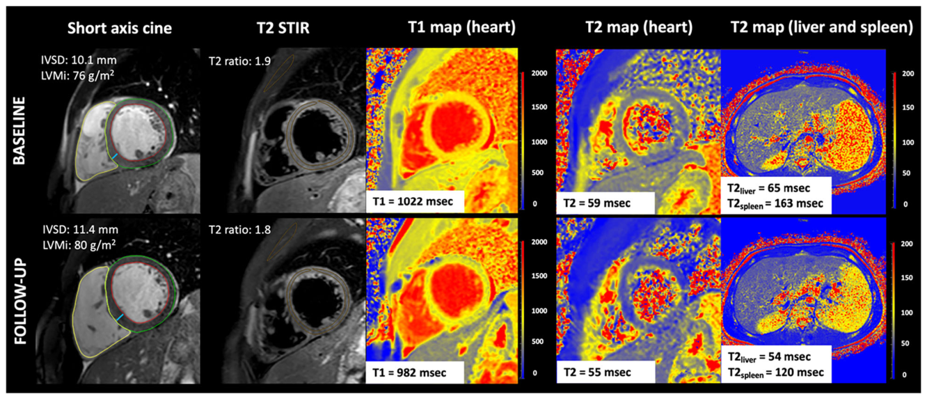
Figure 4. Fig. 4 Clinical example of a 32-year-old man with cryptogenic liver cirrhosis (Child–Pugh class B; Model for End-stage Liver Disease (MELD) score 7) who underwent baseline MRI 7 days prior to TIPS insertion and follow-up MRI 12 months thereafter. Short-axis cine imaging demonstrates an increase in interventricular septal thickness at diastole (IVSD) and a mild increase in left ventricular mass index (LVMi) at follow-up, consistent with structural adaptation to chronic volume loading. T2-weighted short-tau inversion recovery (STIR) imaging shows no focal myocardial edema at baseline or follow-up. In contrast, quantitative myocardial tissue characterization reveals marked reductions in both T1 and T2 relaxation times from elevated baseline values into the normal range at follow-up (center-specific cutoffs: T1 ≥1000 msec; T2 ≥56 msec), indicating resolution of diffuse myocardial edema present prior to TIPS insertion. Corresponding abdominal T2 mapping demonstrates concordant reductions in hepatic and splenic relaxation times, paralleling myocardial tissue normalization and supporting a close association between portal hemodynamics and multiorgan tissue water content解説: 潜因性肝硬変の32歳男性の臨床例を示します。TIPS術前と術後12ヶ月の心臓MRIおよび肝臓・脾臓MRIの画像です。心室中隔厚の増加、左心室心筋量指数の軽度増加、T2 STIR画像での局所的心筋浮腫なし、心筋T1/T2緩和時間の正規化、肝臓および脾臓T2マッピングでの緩和時間の減少が示されています。

コメントを残す