どうも、Beyond the Pixelです。小児期の悪性腎腫瘍は小児がん全体の7%を占め、その中でもウィルムス腫瘍が最も一般的であると広く知られています。しかし、この常識は特定の年齢層においては当てはまりません。思春期後半の対象者、特に10歳代後半では、ウィルムス腫瘍よりも腎細胞癌(RCC)の方が多く見られます。さらに、小児に見られるRCCの病理組織学的スペクトルは成人とは異なり、様々なタイプの腫瘍が存在します。専門家がこれらの腫瘍の臨床的、生物学的、病理組織学的特徴、およびそれらが画像所見に与える影響を理解することは、術前鑑別診断において非常に有用な情報を提供するために不可欠です。本記事では、思春期における主要な腎腫瘍の種類と、その画像診断上の特徴について詳しく解説します。
どうも、Beyond the Pixelです。小児期の悪性腎腫瘍は小児がん全体の7%を占め、その中でもウィルムス腫瘍が最も一般的であると広く知られています。しかし、この常識は特定の年齢層においては当てはまりません。思春期後半の対象者、特に10歳代後半では、ウィルムス腫瘍よりも腎細胞癌(RCC)の方が多く見られます。さらに、小児に見られるRCCの病理組織学的スペクトルは成人とは異なり、様々なタイプの腫瘍が存在します。専門家がこれらの腫瘍の臨床的、生物学的、病理組織学的特徴、およびそれらが画像所見に与える影響を理解することは、術前鑑別診断において非常に有用な情報を提供するために不可欠です。本記事では、思春期における主要な腎腫瘍の種類と、その画像診断上の特徴について詳しく解説します。
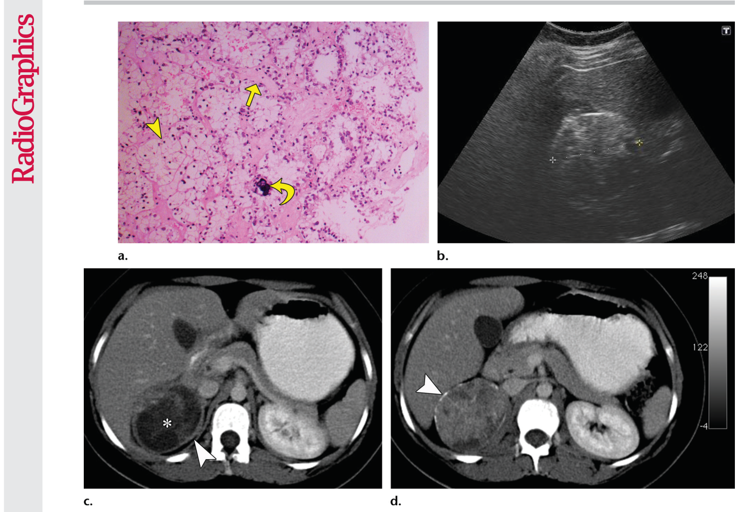
Figure 3. Figure 3. Translocation RCC in a 15-year-old girl. (a) Photomicrograph of a kidney section shows cells with voluminous clear cytoplasm (arrowhead) separated by a fibrovascular network (straight arrow). A psammomatous calcification (curved arrow) also is noted. (Hema toxylin-eosin [H-E] stain; original magnification, 340.) (b) Longitudinal US image shows a mass (calipers) with a hyperechoic rim. (c) Axial iodinated contrast–enhanced CT image shows a fluid-attenuating portion of the mass (*) and a well-defined capsule (arrowhead). (d) Axial CT image obtained at a level slightly lower than that in c shows a more solid portion of the mass with a calcific rim (arrowhead).解説: 15歳の女性に見られる転座型RCC。組織写真では豊富な明澄な細胞質を持つ細胞と砂粒小体が示され(a)、長軸方向のUS画像では高エコー性の辺縁を持つ腫瘤が描出され(b)、造影CTでは液体減衰部分と明瞭な被膜が見られます(c, d)。
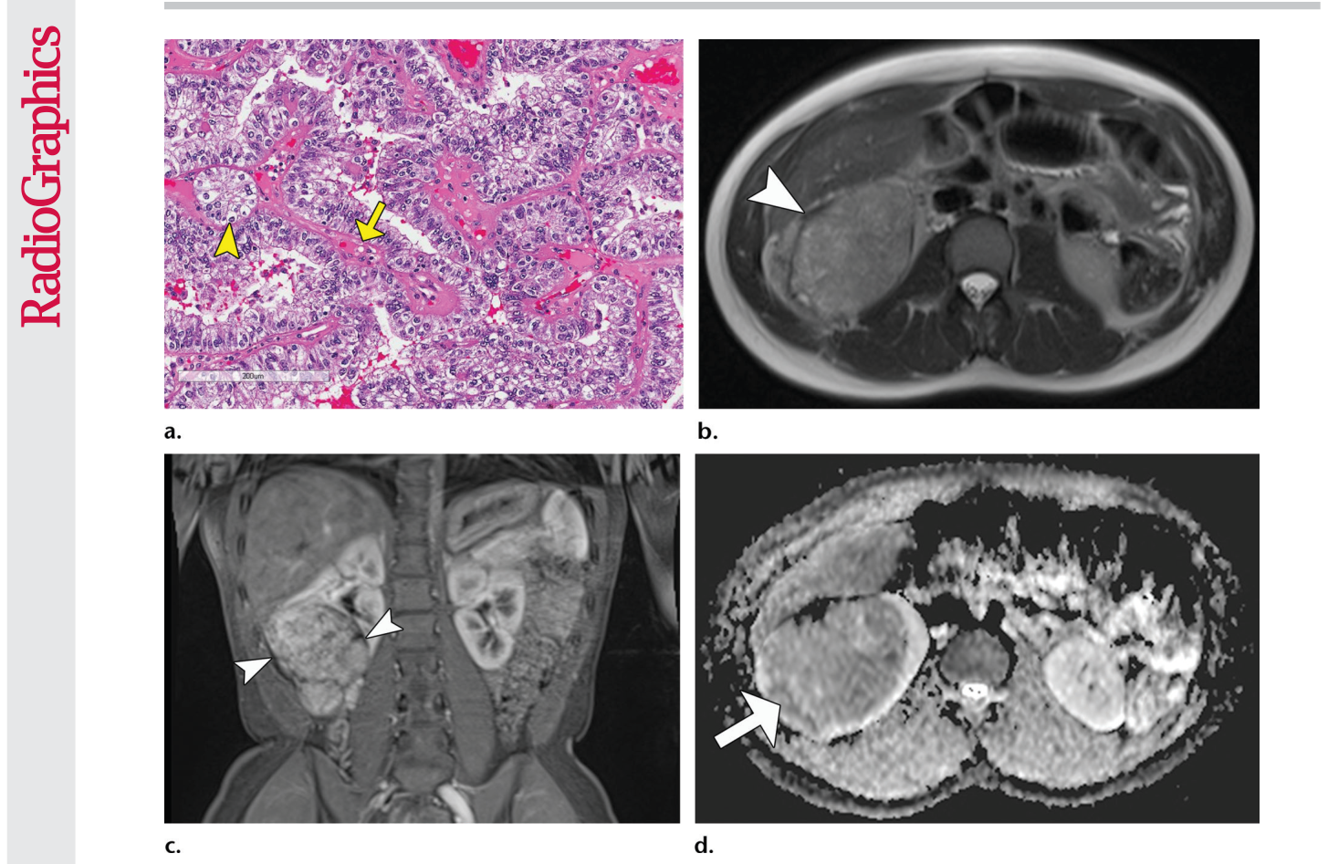
Figure 4. Figure 4. Translocation RCC in a 16-year-old girl. (a) Photomicrograph of a kidney section shows cells with abundant clear to mildly eosinophilic cytoplasm (arrowhead) in nests and papillary arrangements surrounding a fibrovascular core (arrow). (H-E stain; original magnification, 320.) (b) Axial T2-weighted MR image shows an isointense mass with a dark rim (arrowhead), which represents the fibrous capsule. (c) Coronal gadolinium-enhanced fat-saturated T1-weighted MR image shows a het erogeneously enhancing mass with a dark rim (arrowheads). (d) Axial apparent diffusion coefficient map from an echo-planar diffusion-weighted sequence shows restricted diffusion (arrow), consistent with a densely cellular tumor.解説: 16歳の女性に見られる転座型RCC。組織写真では豊富な明澄から軽度好酸性の細胞質を持つ細胞が乳頭状に配列しており(a)、軸方向T2強調MR画像では暗い辺縁を伴う等信号の腫瘤が示され(b)、冠状ガドリニウム造影T1強調脂肪抑制MR画像では不均一に造影される腫瘤と暗い辺縁が見られ(c)、エコープレナー拡散強調シーケンスからのADCマップでは高細胞密度腫瘍と一致する拡散制限が示されています(d)。
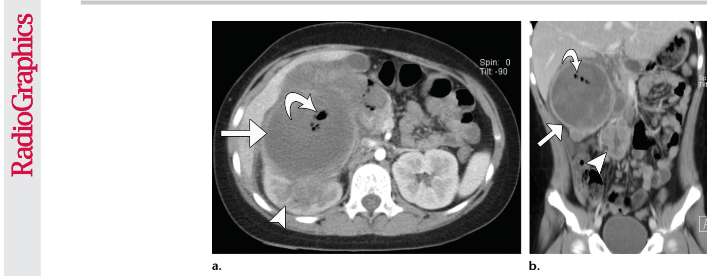
Figure 5. Figure 5. Papillary RCC in a 10-year-old boy with sepsis. (a) Axial iodinated contrast–enhanced CT im age shows a mass (straight arrow) centered in the right kidney (arrowhead) that has lower enhancement than the cortex during the corticomedullary phase. Small collections of air (curved arrow) within the tumor were found at surgery to be caused by a duodenal fistula. (b) Coronal CT image shows the tumor (straight arrow), air (curved arrow), and retroperitoneal nodal metastasis (arrowhead).解説: 敗血症を伴う10歳の男児に見られる乳頭状RCC。造影CT軸位像では、皮質期に皮質よりも造影効果の低い腫瘤が右腎臓の中央に認められ、腫瘍内に少量の空気の集まりが見られます(a)。冠状CT画像では腫瘍、空気、および後腹膜リンパ節転移が確認できます(b)。
孤立性の腎腫瘍がある場合、年齢が鑑別診断を絞り込む上で最も有用な特徴です。ウィルムス腫瘍は48ヶ月未満で診断されることが多く、95%の症例は10歳未満で診断されます。しかし、15~19歳の年齢層では、RCCがウィルムス腫瘍よりも一般的です。小児RCCは通常、ウィルムス腫瘍よりも小さく、石灰化しやすい傾向があります(RCCで最大25% vs ウィルムス腫瘍で最大9%)。腎静脈への浸潤はRCCとウィルムス腫瘍の両方で見られますが、RCCは骨への転移が多いという特徴があります。
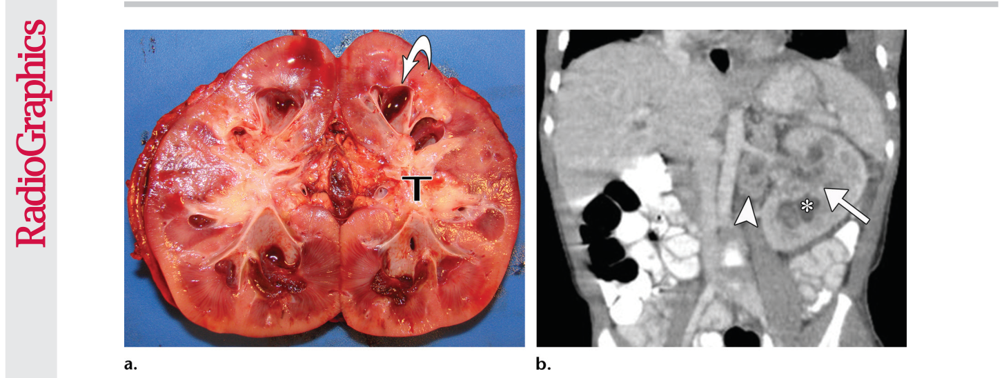
Figure 6. Figure 6. Renal medullary carcinoma in a 9-year-old boy with gross hematuria, who was treated for a urinary tract infection and later discovered to have the sickle cell trait. (a) Bivalved gross kidney specimen shows a central yellow- tan tumor (T) that has ill-defined margins and extends into the renal sinus. Dilated calyces (arrow) also are noted. (b) Coronal iodinated contrast–enhanced CT image shows an ill-defined mass (arrow) in the left kidney, with the re niform shape preserved; an enlarged heterogeneous retroperitoneal lymph node (arrowhead); and a dilated calyx (*).解説: 肉眼的血尿を呈し、尿路感染症の治療を受け、後に鎌状赤血球形質を持つことが判明した9歳の男児に見られる腎髄様癌。割面された肉眼的腎臓検体では、境界不明瞭で腎洞に広がる中心性の黄色褐色腫瘍と拡張した腎杯が示されています(a)。冠状造影CT画像では、左腎臓に境界不明瞭な腫瘤があり、腎臓の形態は保たれており、腫大した不均一な後腹膜リンパ節と拡張した腎杯が見られます(b)。
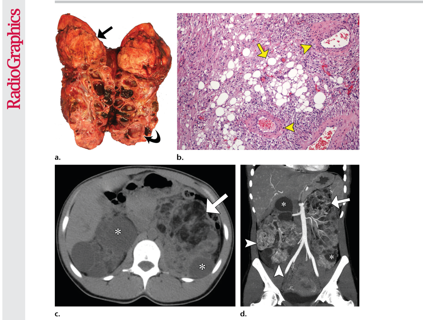
Figure 9. Figure 9. Angiomyolipomas in a 23-year-old woman with tuberous sclerosis. (a) Bivalved gross kidney specimen shows a yellow-tan mass (straight arrow) in the upper pole and numerous cysts (curved arrow). (b) Photomicrograph of a kidney section shows fat cells (arrow) and vessels with thick walls (arrowheads) within the tumor. (H-E stain; original magnification, 310.) (c) Axial nonenhanced CT image shows a heterogeneous mass with fat attenuation (arrow), as well as multiple bilateral fluid-attenuating cysts (*). (d) Coronal image reformatted from CT angiography shows the enhancing components of the fat-con taining tumor (arrow), as well as additional smaller enhancing tumors (arrowheads). Nonenhancing cysts (*) also are seen. Also noted are numerous small, presumed flash-filling hemangiomas in the liver.解説: 結節性硬化症の23歳の女性に見られる血管筋脂肪腫。割面された肉眼的腎臓検体では、上極に黄色褐色の腫瘤と多数の嚢胞が示されています(a)。組織写真では腫瘍内に脂肪細胞と厚い壁を持つ血管が見られます(b)。軸方向非造影CT画像では、脂肪減衰を持つ不均一な腫瘤と多発性の両側性液体減衰嚢胞が示されています(c)。CT血管造影の再構成冠状画像では、脂肪含有腫瘍の造影される成分と追加の小さな造影腫瘍、および非造影嚢胞が示されています(d)。
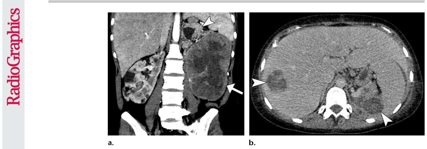
Figure 10. Figure 10. Metastatic perivascular epithelioid cell tumor in a 20-year-old woman with TSC. (a) Coronal contrast- enhanced reformatted CT image shows a large mass (arrow) with central fluid attenuation that corresponded to old hemorrhage at gross specimen inspection. No hypoattenuating fat is identified in the tumor. Linear high- attenuating calcification, bilateral fat-containing angiomyolipomas (arrowhead), and renal cysts also are seen. (b) Axial CT image obtained after resection shows hypoattenuating masses (arrowheads) in the liver and left renal bed, which were confirmed at biopsy to represent metastasis and recurrent tumor. Chest CT images (not shown) showed multiple large nodules representing metastases.解説: TSCの20歳の女性に見られる転移性血管周囲上皮様細胞腫瘍。冠状造影再構成CT画像では、中心に液体減衰を示す大きな腫瘤が示されており、これは肉眼検体検査で古い出血に対応していました。腫瘍内には低減衰の脂肪は確認されず、線状の高減衰性石灰化、両側性の脂肪含有血管筋脂肪腫、および腎嚢胞も確認できます(a)。切除後に撮影された軸方向CT画像では、肝臓および左腎床に低減衰性の腫瘤が示されており、生検で転移および再発腫瘍と確認されました(b)。
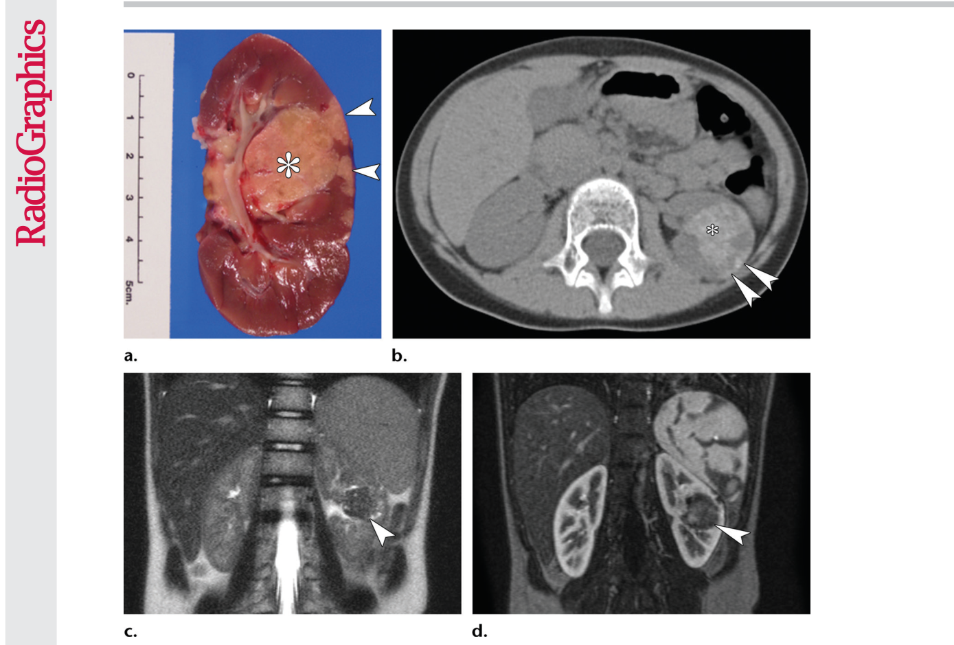
Figure 11. Figure 11. Metanephric adenoma in a girl. (a) Sectioned gross kidney specimen shows a central yellow- tan circumscribed mass (*) with satellite nodules (arrowheads) that extend to the capsule. (b) Axial nonenhanced CT image shows a hyperattenuating mass (*) and the peripheral nodules (arrowheads). (c) Coronal T2-weighted MR image shows a central mass (arrowhead) that is hypointense compared with the renal cortex and medulla, suggesting calcification or a densely cellular tumor. (d) Coronal arte rial phase MR angiogram shows the mass (arrowhead) with lower enhancement than the renal cortex.解説: 女児に見られるメタネフリック腺腫。割面された肉眼的腎臓検体では、中心に黄色褐色の境界明瞭な腫瘤と被膜に広がる衛星結節が示されています(a)。軸方向非造影CT画像では、高吸収域の腫瘤と末梢結節が示されています(b)。冠状T2強調MR画像では、腎皮質および髄質と比較して低信号の中心性腫瘤が示されており、石灰化または高細胞密度腫瘍が示唆されます(c)。冠状動脈相MRアンギオグラムでは、腎皮質と比較して造影効果の低い腫瘤が示されています(d)。
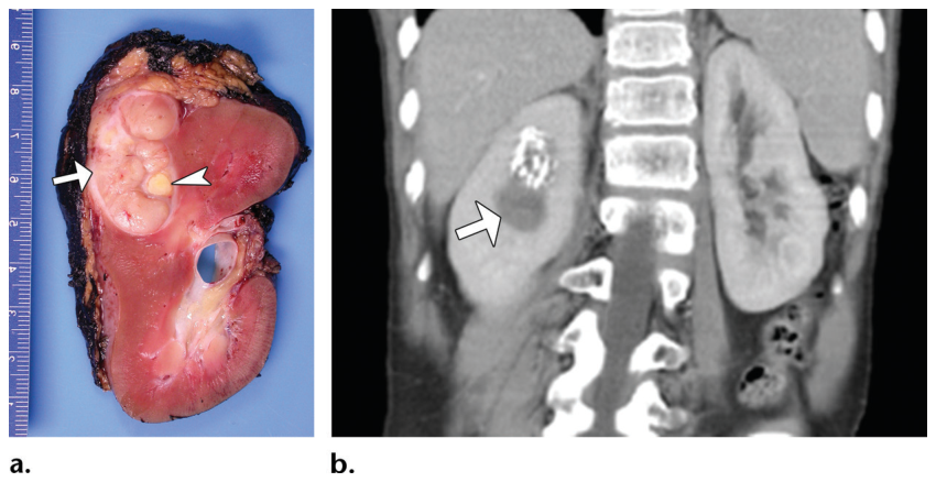
Figure 12. Figure 12. Metanephric adenoma in a 7-year-old boy. (a) Sectioned gross specimen shows a well-circumscribed lobulated white-gray tumor (arrow) with a focal area of necrosis (arrowhead). (b) Coronal iodinated contrast–enhanced reformatted CT image shows a circumscribed hypoenhancing mass (arrow) adjacent to a cluster of coarse calcifications.解説: 7歳の男児に見られるメタネフリック腺腫。割面された肉眼的検体では、境界明瞭な小葉状の白灰色の腫瘍と局所的な壊死領域が示されています(a)。冠状造影再構成CT画像では、粗大な石灰化の集まりに隣接する境界明瞭な低造影腫瘤が示されています(b)。
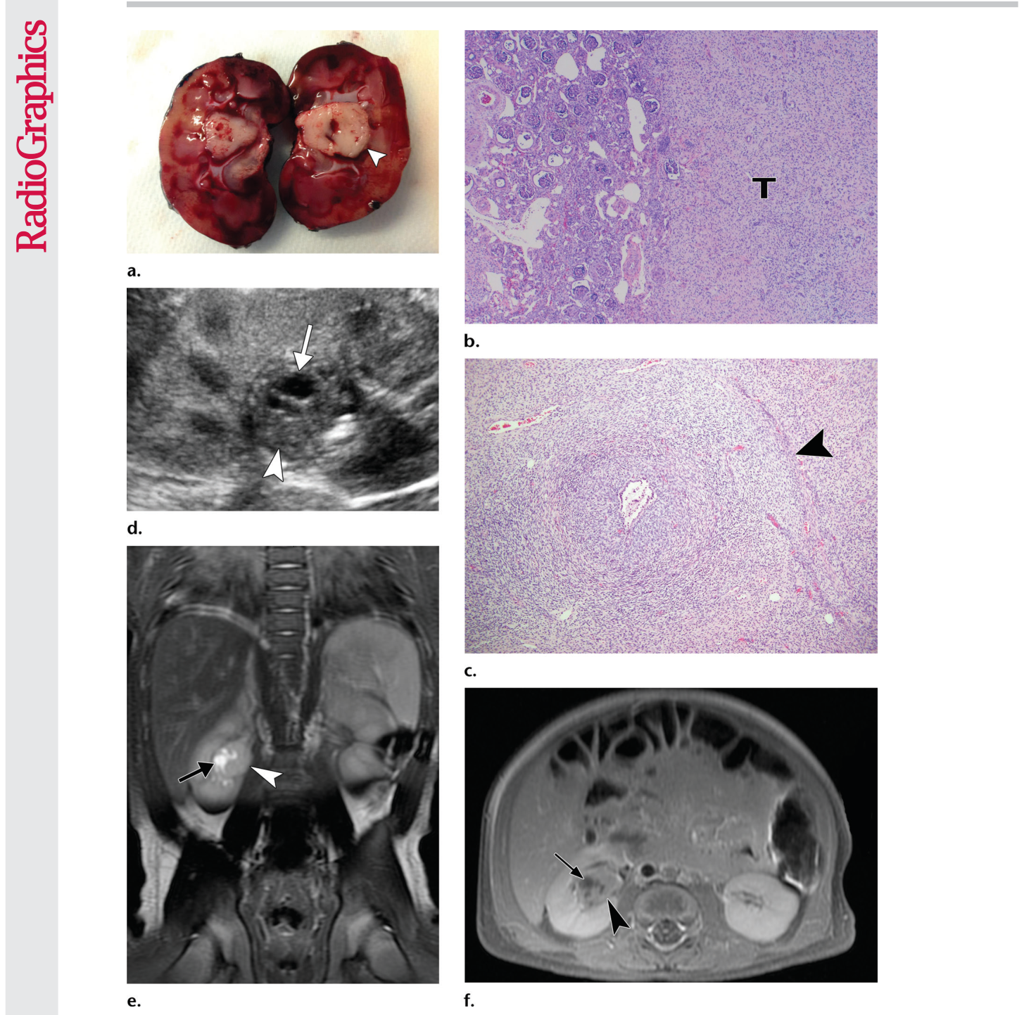
Figure 14. Figure 14. Metanephric stromal tumor. (a) Bivalved gross kidney specimen from a 3-month-old boy shows a well-defined tan-gray tumor (arrowhead) that is centered in the renal medulla and has small areas of hemor rhage. (b) Photomicrograph of a kidney section from a 2-month-old boy shows a nonencapsulated spindle cell tumor (T) on the right and the adjacent kidney on the left. (H-E stain; original magnification, 32.) (c) Photomi crograph of a kidney section from the same boy shows the “onion-skin” or collarette appearance of concentric layers of tumor cells (arrowhead) surrounding an entrapped tubule. (H-E stain; original magnification, 310.) (d) Longitudinal US image in the boy in a shows a hilar mass (arrowhead) with anechoic cystic components (arrow). (e) Coronal T2-weighted MR image in the boy in a shows a hilar mass (arrowhead) that is isoin tense compared with the renal parenchyma. There are small fluid–signal-intensity cysts (arrow) within the mass. (f) Axial gadolinium-enhanced T1-weighted fat-saturated MR image in the boy in a shows the mass (arrowhead) with lower enhancement compared with the renal parenchyma. The fluid-filled cysts (arrow) are not enhanced.解説: メタネフリック間質性腫瘍。3ヶ月男児の割面された肉眼的腎臓検体では、腎髄質に中心を持つ境界明瞭な黄褐色灰色の腫瘤と小さな出血領域が示されています(a)。2ヶ月男児の腎臓組織写真では、非被包性の紡錘細胞腫瘍と隣接する腎臓、およびその間に炎症反応が見られます(b)。同じ男児の腎臓組織写真では、「玉ねぎの皮」または捕捉された尿細管を囲む腫瘍細胞の同心円状の層である襟状の外観が示されています(c)。3ヶ月男児の長軸方向US画像では、無エコー性嚢胞成分を持つ門部腫瘤が示されています(d)。同じ男児の冠状T2強調MR画像では、腎実質と比較して等信号の門部腫瘤と腫瘤内に小さな液体信号強度の嚢胞が見られます(e)。同じ男児の軸方向ガドリニウム造影T1強調脂肪抑制MR画像では、腎実質と比較して造影効果の低い腫瘤と造影されない液体充満嚢胞が示されています(f)。
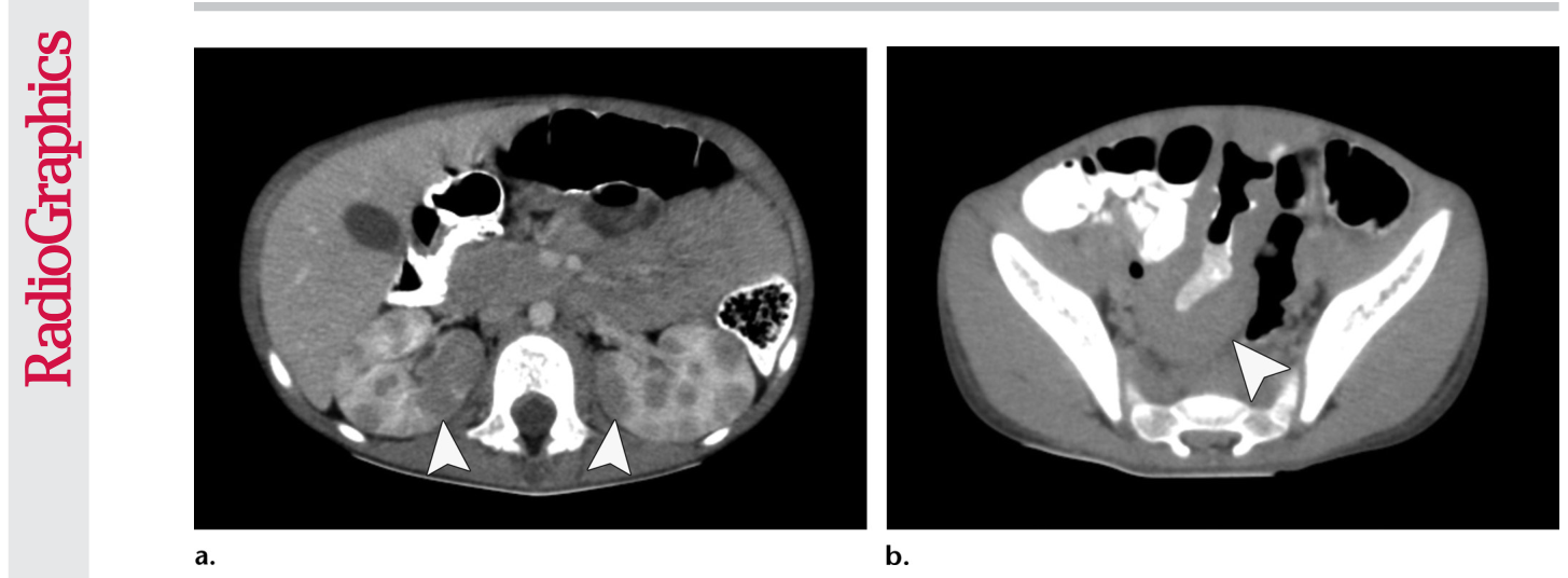
Figure 16. Figure 16. Burkitt lymphoma in a 5-year-old boy. (a) Axial contrast-enhanced CT image shows multiple hypoattenuating circum scribed masses (arrowheads) in both kidneys. (b) Axial CT image of the pelvis shows diffuse wall thickening of the ileum (arrowhead), which is typical of Burkitt lymphoma.解説: 5歳の男児に見られるバーキットリンパ腫。軸方向造影CT画像では、両腎臓に多発性の低減衰性境界明瞭腫瘤が示されています(a)。骨盤の軸方向CT画像では、バーキットリンパ腫に典型的な回腸のびまん性壁肥厚が示されています(b)。
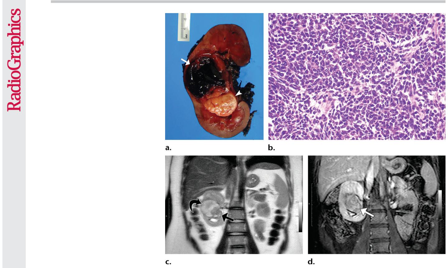
Figure 17. Figure 17. PNET in a 15-year-old girl. (a) Sectioned gross kidney specimen shows a central tan-white lobular tumor (arrowhead) with small areas of hemorrhage and an adjacent intrapa renchymal hematoma (arrow). (b) Photomicrograph of a kidney section demonstrates sheets of small round cells with scant cytoplasm and coarse chromatin. (H-E stain; original magnifica tion, 340.) (c) Coronal T2-weighted MR image shows a hypointense central mass (straight arrow) consistent with a densely cellular tumor. The hyperintense component (curved arrow) adjacent to the tumor represents the hematoma. (d) Coronal gadolinium-enhanced MR im age shows the mass (arrow) enhanced but less intensely compared with the adjacent renal parenchyma.解説: 15歳の女児に見られるPNET。割面された肉眼的腎臓検体では、中心に黄白色の小葉状腫瘍と小さな出血領域、および隣接する腎実質内血腫が示されています(a)。腎臓組織写真では、細胞質が少なく粗大なクロマチンを持つ小型円形細胞のシートが描出されています(b)。冠状T2強調MR画像では、高細胞密度腫瘍と一致する低信号の中心性腫瘤が示されています。腫瘍に隣接する高信号成分は血腫を表しています(c)。冠状ガドリニウム造影MR画像では、腫瘤は造影されますが、隣接する腎実質と比較して造影効果は弱いです(d)。
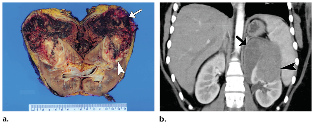
Figure 18. Figure 18. Renal PNET in a 16-year-old girl. (a) Bisected gross kidney specimen shows a tan-brown variegated hemorrhagic mass (arrowhead) with an adjacent subcapsular hemorrhage (arrow). (b) Coronal iodinated contrast–enhanced reformatted CT image shows the upper pole mass (arrowhead) with lower enhancement compared with the adjacent renal cortex during the corticomedullary phase. The subcap sular fluid-attenuation collection (arrow) corresponds to the chronic hemorrhage seen at gross inspection.解説: 16歳の女児に見られる腎PNET。二分された肉眼的腎臓検体では、黄褐色で多様な出血性腫瘤と隣接する被膜下出血が示されています(a)。冠状造影再構成CT画像では、皮質髄質相において隣接する腎皮質と比較して造影効果の低い上極腫瘤と、肉眼検査で見られた慢性出血に対応する被膜下液体減衰性病変が示されています(b)。
Figure 19. Figure 19. PNET in a 17-year-old girl. (a) Axial delayed phase contrast-enhanced CT image shows a central mass (arrowhead) enhancing less intensely compared with the adjacent parenchyma and extension of the tumor through the renal vein (straight arrow) into the inferior vena cava. A small enhancing mass (curved arrow) also is seen in the spinal canal. (b) Axial CT image at the level of the diaphragm shows a filling defect (arrow) in the intrahepatic inferior vena cava and a left pulmonary metastatic nodule (arrowhead).


コメントを残す