どうも、Beyond the Pixelです。妊娠中や授乳期における乳房の生理的変化は、乳がんのスクリーニングと診断に特有の課題をもたらします。この時期は乳腺組織の密度が増加し、乳房のサイズも変化するため、臨床的な触診やマンモグラフィによる所見の評価が難しくなることがあります。しかし、妊娠関連乳がん(PABC)の診断が遅れると、予後不良につながる可能性があるため、画像評価を遅らせるべきではありません。本記事では、妊娠中および授乳期における乳房の画像診断について、安全かつ効果的なアプローチと、遭遇しやすいさまざまな乳房の異常について、最新の論文に基づいて詳しく解説します。
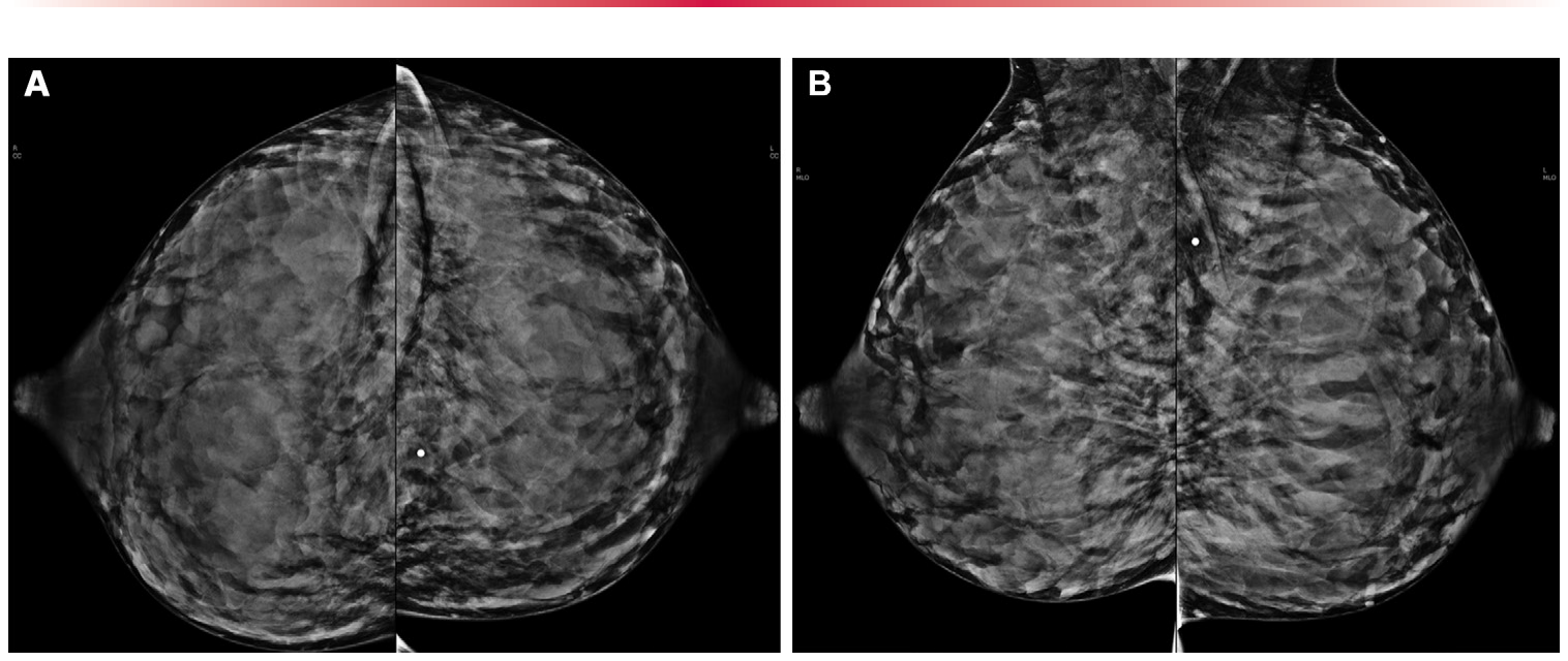
Figure 1. Figure 1. Normal mammographic appearance of the breasts in a 33-year-old lactating woman who presented with a palpable abnormality in the left breast. Bilateral craniocaudal (A) and mediolateral oblique (B) mammograms show symmetric increased parenchymal density (breast composition category D, ex- tremely dense). No underlying mammographic abnormality is identified. The palpable area of concern corresponded with normal breast tissue on US images (not shown).解説: 33歳の授乳中の女性の乳房の正常なマンモグラフィ画像を示します。左乳房に触知可能な異常を訴え来院しました。両側頭尾方向(A)および内外斜位方向(B)のマンモグラムは、対称的な乳腺密度の増加(乳腺構成カテゴリーD、極めて高密度)を示しています。マンモグラフィ上の異常は認められず、触知部位は超音波検査で正常な乳腺組織に対応していました。
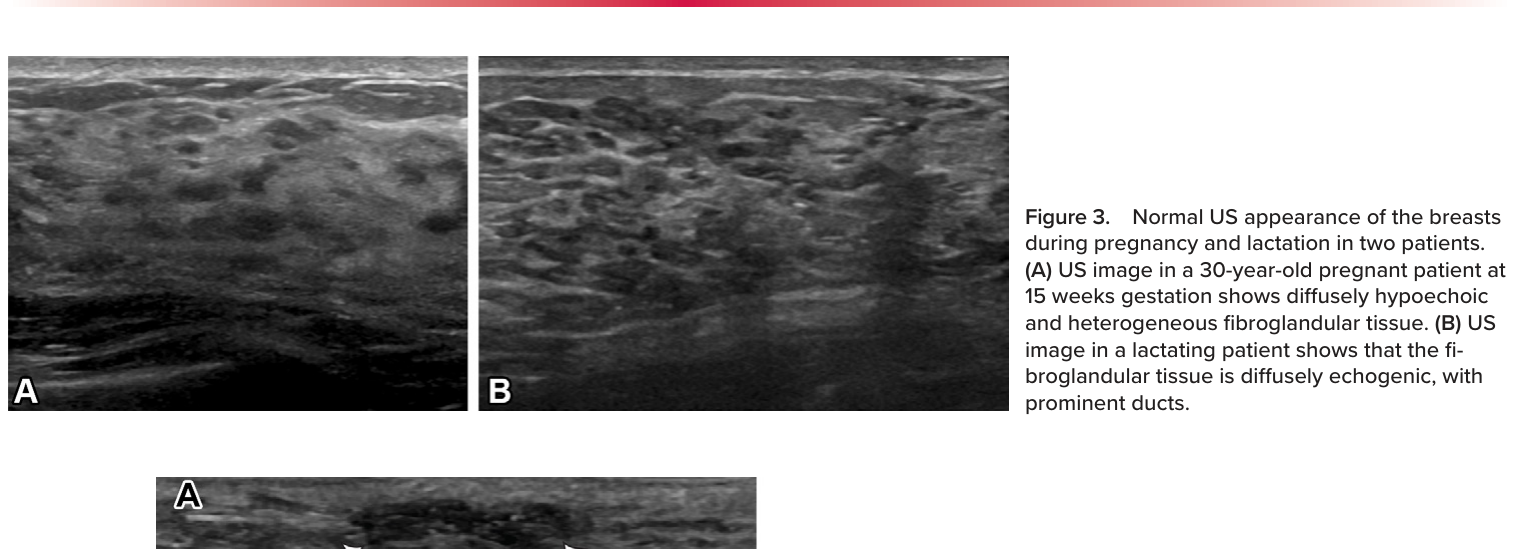
Figure 4. Figure 4. Lactational changes in a 33-year-old lactating woman who presented with a palpable abnormality in the left breast. Mammogram (not shown) showed no underlying abnormality. (A) US image shows an oval hypoechoic mass with indistinct margins (arrows), which correlated with the palpable mass reported by the patient. The mass was cate- gorized as BI-RADS 4A, and US-guided biopsy was subse- quently performed. (B) Photomicrographs show cores with variably sized acini (left) and variably dilated acinar units (, right) in lobules containing intraluminal secretions consistent with lactational changes. Note preservation of outer myoep- ithelial layers (arrowheads, right). (Hematoxylin-eosin stain; original magnification, left, 4; right, 200.)解説: この画像は論文のFigure 3であり、妊娠中および授乳中の2名の対象者の乳房の正常な超音波(US)画像を示しています。(A) 妊娠15週の30歳の妊婦におけるUS画像では、びまん性に低エコーで不均一な線維腺組織が確認できます。(B) 授乳中の対象者におけるUS画像では、線維腺組織がびまん性に高エコーで、乳管が目立つ状態が示されています。これは指定されたFigure 4のキャプション(授乳変化を示す画像)とは一致しません。⚠ 自動抽出画像の検証で一致を確認できませんでした。正確な内容は元論文のFigure 4をご参照ください。
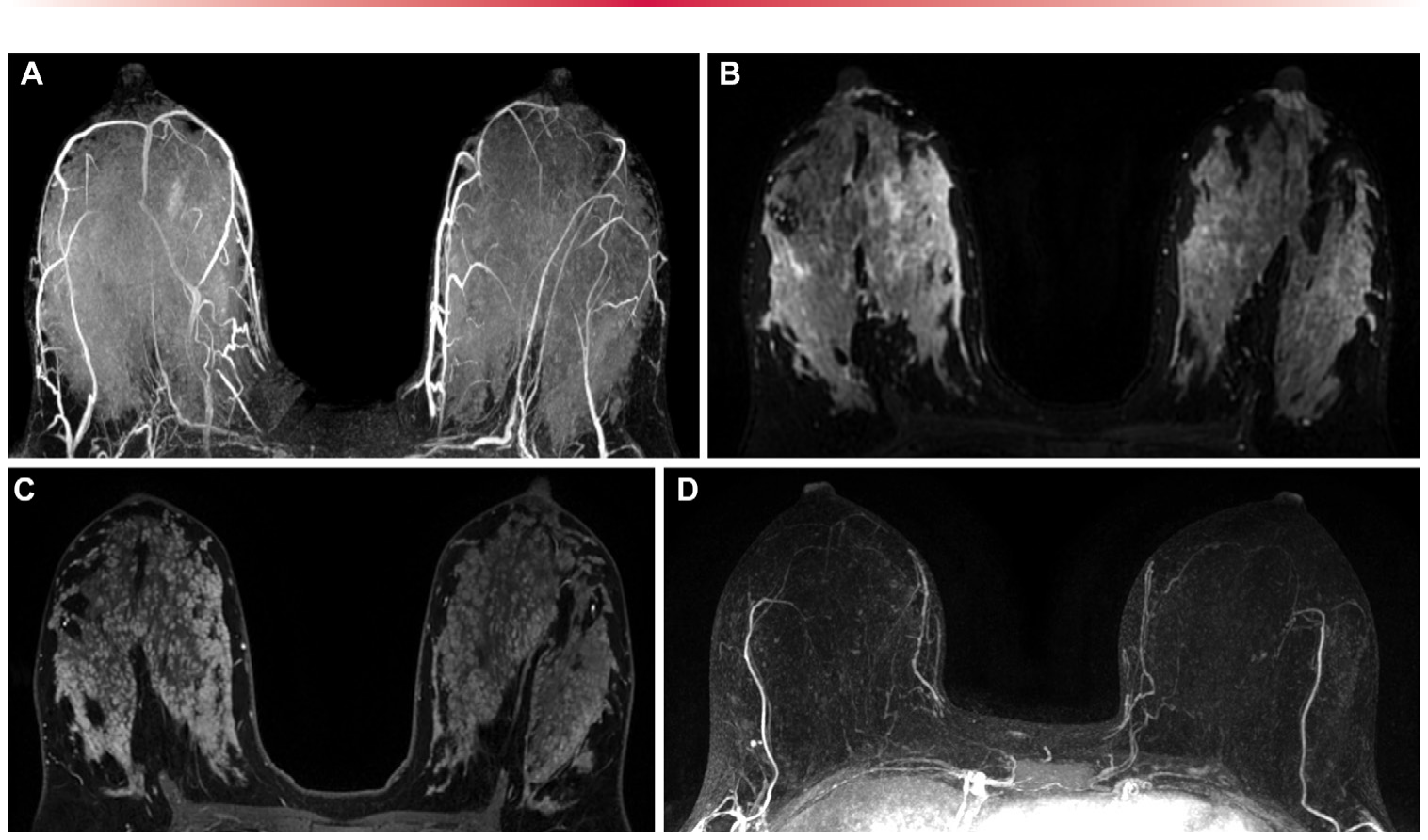
Figure 5. Figure 5. Normal MRI appearance of the breasts in a high-risk 36-year-old lactating woman with a family history of breast cancer. (A) Axial maximum intensity projection MR image shows marked bilateral background parenchymal enhancement and increased vascularity. (B) Axial fat-saturated T2-weighted MR image shows diffuse increased signal intensity. (C) Axial fat-saturated early phase contrast-enhanced MR image shows an increased amount of fibroglandular tissue. (D) Maximum intensity projection MR image acquired in the patient before pregnancy and lactation shows only mild background parenchymal enhancement.解説: 乳がんの家族歴を持つ高リスクの36歳の授乳中の女性の乳房の正常なMRI画像を示します。(A) 軸位最大強度投影(MIP)MR画像は、著しい両側性背景実質造影増強と血管増加を示しています。(B) 軸位脂肪抑制T2強調MR画像は、びまん性の信号強度増加を示しています。(C) 軸位脂肪抑制早期造影MR画像は、線維腺組織の量の増加を示しています。(D) 妊娠・授乳前の同対象者のMIP MR画像は、軽度の背景実質造影増強のみを示しています。
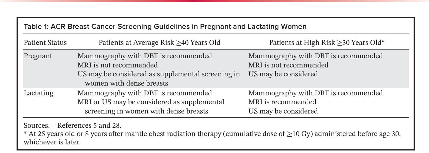
Table 1. Table 1: ACR Breast Cancer Screening Guidelines in Pregnant and Lactating Women解説: ACR(米国放射線専門家会)が定める妊娠中および授乳中の女性における乳がんスクリーニングガイドラインを要約した表です。平均リスクの40歳以上の対象者と高リスクの30歳以上の対象者に対し、妊娠中と授乳中のそれぞれで推奨されるマンモグラフィ、MRI、USなどのスクリーニング方法が示されています。
Figure 6. Figure 6. Diagnostic imag- ing algorithm for pregnant and lactating women with a new clinical concern. For pregnant patients or pa- tients younger than age 30 who are breastfeeding with a BI-RADS category 4C or 5 abnormality, mammography ideally should be performed before biopsy.解説: この画像は論文のTable 1であり、ACR(米国放射線専門家会)が定める妊娠中および授乳中の女性における乳がんスクリーニングガイドラインを要約したものです。この表には、平均リスクの40歳以上の対象者と高リスクの30歳以上の対象者に対し、妊娠中と授乳中のそれぞれで推奨されるマンモグラフィ、MRI、USなどのスクリーニング方法が示されています。これは指定されたFigure 6のキャプション(診断画像アルゴリズム)とは一致しません。⚠ 自動抽出画像の検証で一致を確認できませんでした。正確な内容は元論文のFigure 6をご参照ください。
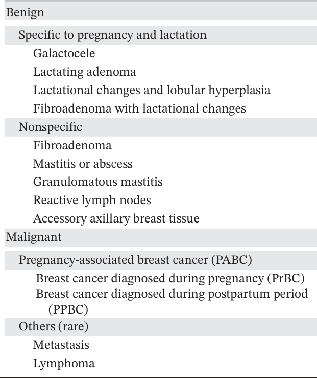
Table 2. Table 2: Breast Abnormalities Related to Pregnancy and Lactation解説: 妊娠中および授乳期に関連する乳房の異常をリストアップした表です。妊娠・授乳期に特有の良性病変(乳瘤、授乳腺腫など)と、非特異的な良性病変(線維腺腫、乳腺炎など)、そして悪性病変(妊娠関連乳がん、転移、リンパ腫)に分類されています。
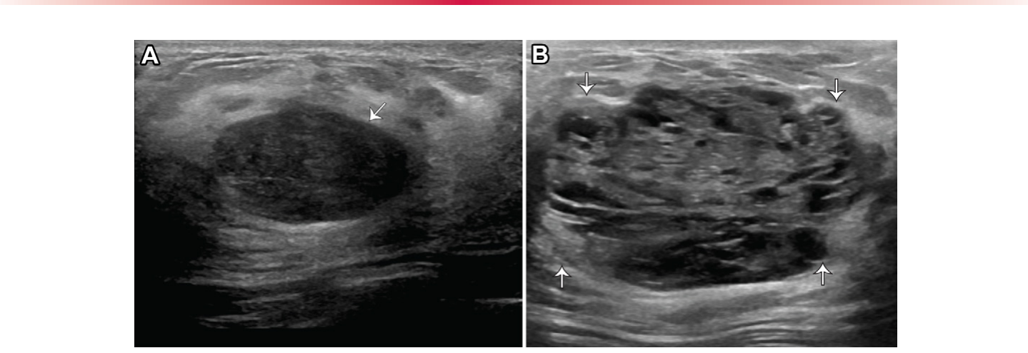
Figure 11. Figure 11. Fibroadenoma with lactational changes in a 32-year-old woman who presented with a lump in her left breast. (A) US image acquired at 15 weeks gestation shows an oval hypoechoic mass with circumscribed margins (arrow) and posterior acoustic enhancement, which was categorized as BI-RADS 3, with short-interval follow-up recommended. (B) Follow-up postpartum US image acquired while the patient was breastfeeding shows an inter- val increase in the size and heterogeneity of the mass (arrows). The mass was recategorized as BI-RADS 4A, and subsequent US-guided biopsy results confirmed the diagnosis of a fibroadenoma with lactational changes.解説: 左乳房にしこりを訴えて来院した32歳の女性における、授乳変化を伴う線維腺腫の超音波画像を示します。(A) 妊娠15週目に撮影されたUS画像では、境界明瞭な楕円形の低エコー腫瘤(矢印)と後方音響増強が認められ、BI-RADS 3と分類され短期間の経過観察が推奨されました。(B) 出産後の授乳中に撮影されたフォローアップUS画像では、腫瘤のサイズが増加し、不均一性が増している(矢印)ことが確認できます。この腫瘤はBI-RADS 4Aに再分類され、その後のUSガイド下生検で授乳変化を伴う線維腺腫と診断されました。
、
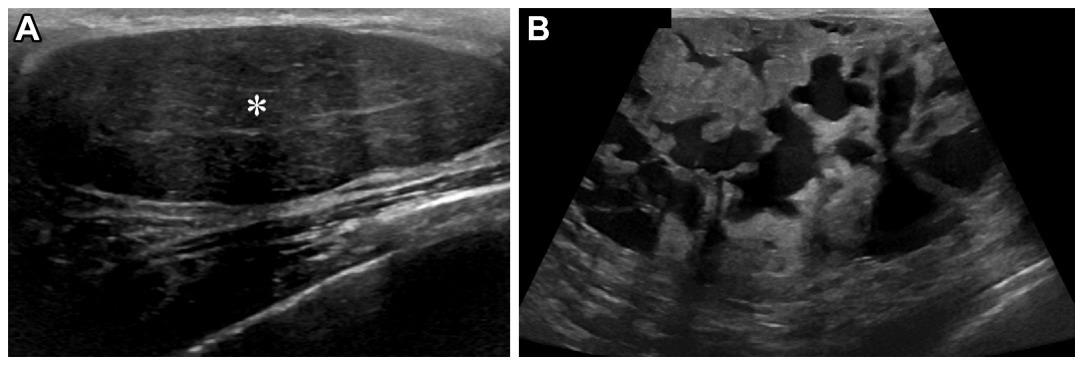
Figure 12. Figure 12. Fibroadenoma with lactational changes in a 34-year-old lactating woman who presented with an en- larging right breast lump at the site of biopsy-proven fibroadenoma. (A) US image acquired 4 years earlier when the patient was not lactating shows an oval hypoechoic mass with circumscribed margins (*). US-guided biopsy (not shown) showed a benign fibroadenoma. (B) US image acquired at presentation when the patient was lac- tating shows the substantially increased size of the mass, with new internal cystic spaces and indistinct margins. Due to the complex cystic and solid mass appearance and interval growth of the mass, excisional biopsy was ulti- mately performed, and the result confirmed the diagnosis of a fibroadenoma with lactational changes.解説: 生検で線維腺腫と診断された部位に増大する右乳房のしこりを訴えて来院した34歳の授乳中の女性における、授乳変化を伴う線維腺腫の超音波画像を示します。(A) 4年前に非授乳期に撮影されたUS画像では、境界明瞭な楕円形の低エコー腫瘤(アスタリスク)が示されています。USガイド下生検で良性線維腺腫と診断されました。(B) 来院時(授乳中)に撮影されたUS画像では、腫瘤が著しく増大し、新たな内部嚢胞性空間と不明瞭な境界が認められます。この複雑な嚢胞性・固形腫瘤様の外観と腫瘤の増大のため、最終的に切除生検が行われ、授乳変化を伴う線維腺腫の診断が確定されました。
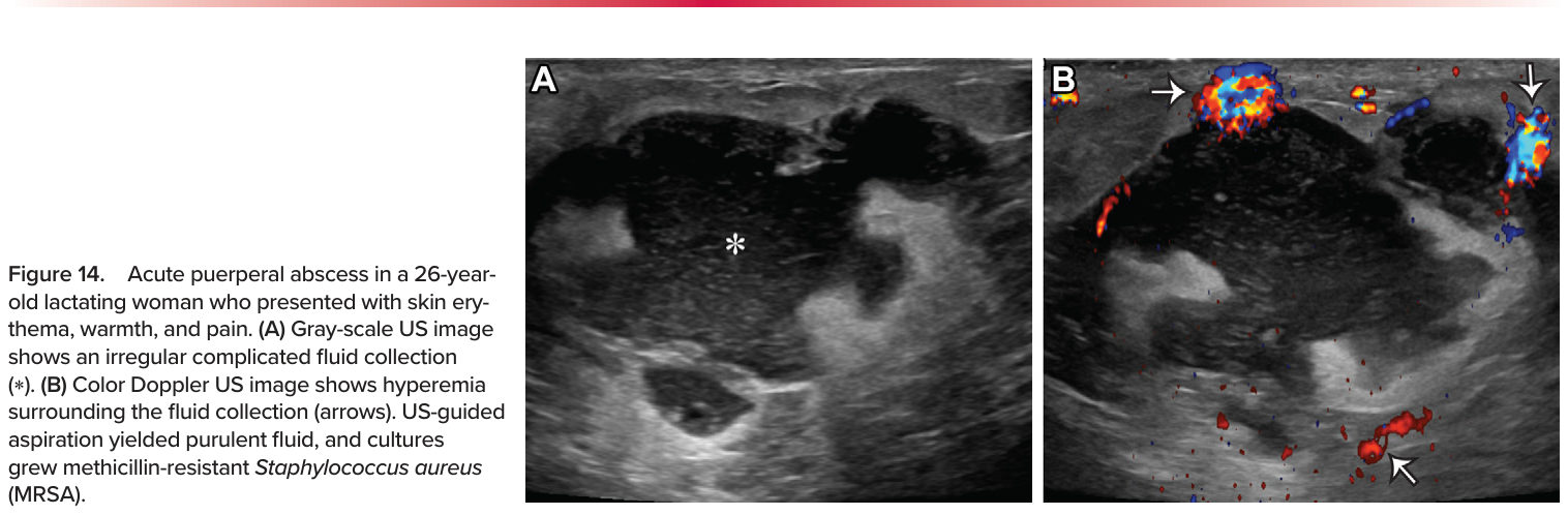
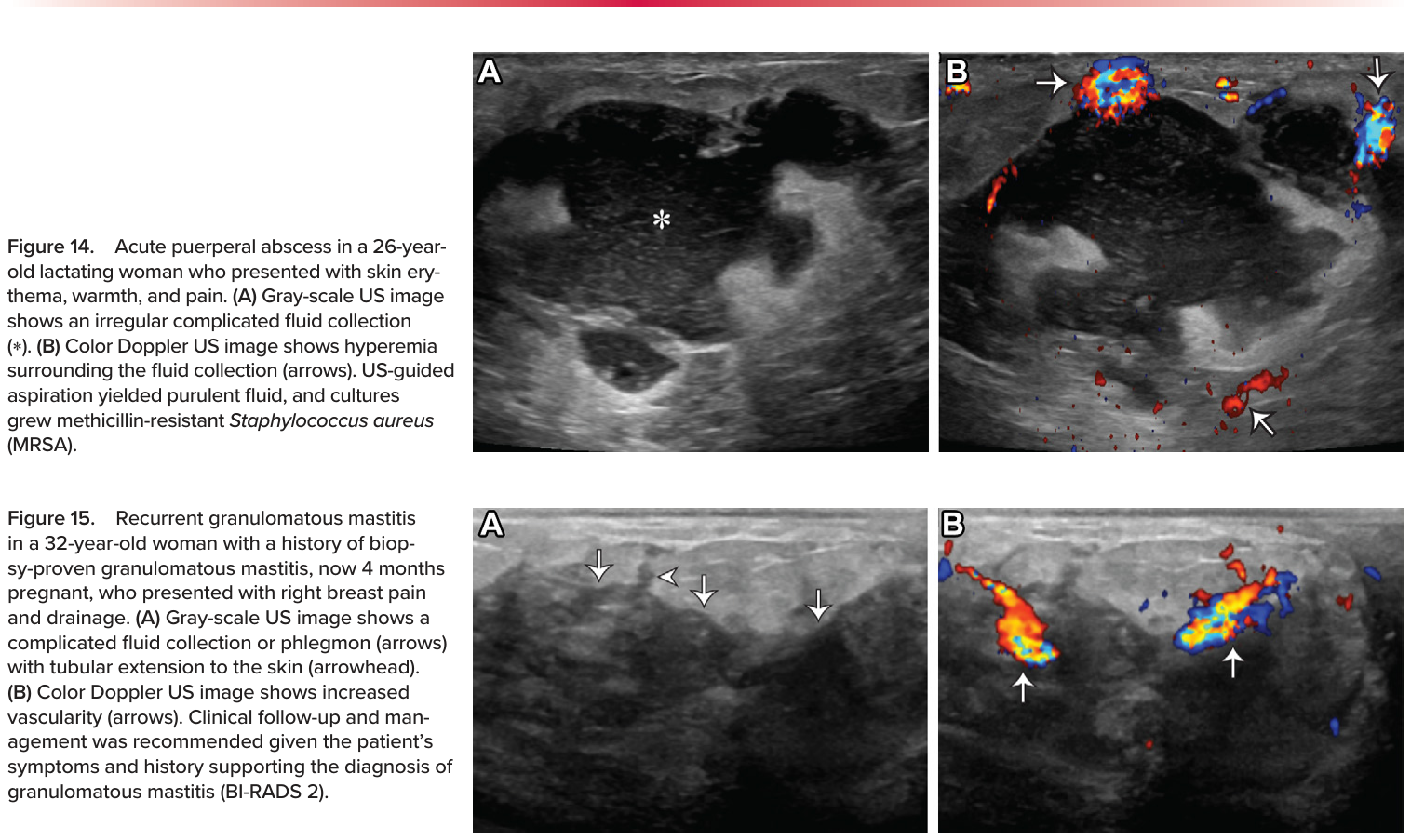
Figure 15. Figure 15. Recurrent granulomatous mastitis in a 32-year-old woman with a history of biop- sy-proven granulomatous mastitis, now 4 months pregnant, who presented with right breast pain and drainage. (A) Gray-scale US image shows a complicated fluid collection or phlegmon (arrows) with tubular extension to the skin (arrowhead). (B) Color Doppler US image shows increased vascularity (arrows). Clinical follow-up and man- agement was recommended given the patient’s symptoms and history supporting the diagnosis of granulomatous mastitis (BI-RADS 2).解説: この画像は論文のFigure 14であり、皮膚紅斑、熱感、痛みを訴えて来院した26歳の授乳中の女性における急性産褥期膿瘍の超音波画像を示しています。(A) グレースケールUS画像は、不規則な複雑な液体貯留(アスタリスク)を示しています。(B) カラードップラーUS画像は、液体貯留周囲の血流増加(矢印)を示しています。USガイド下吸引で化膿性液体が得られ、培養でメチシリン耐性黄色ブドウ球菌(MRSA)が検出されました。これは指定されたFigure 15のキャプション(再発性肉芽腫性乳腺炎)とは一致しません。⚠ 自動抽出画像の検証で一致を確認できませんでした。正確な内容は元論文のFigure 15をご参照ください。
Figure 16. Figure 16. Benign reactive lymph nodes in a 34-year-old lactating woman who presented with a pal- pable abnormality in the right axilla after recent ipsilateral COVID-19 vaccination. A mammogram was negative for abnormality (not shown). (A) Gray-scale US image shows prominent lymph nodes in the right axilla, with uniform corti- cal thickening measuring up to 6 mm (double-headed arrow). The morphology of the lymph nodes is otherwise normal, with preserved fatty hila. (B) Color Doppler US image shows hilar vascularity (arrow). The findings are consistent with reactive lymphade- nopathy due to recent ipsilateral vaccination (BI-RADS 2). The patient received clinical follow-up, with no palpable abnormality on physical examination.解説: この画像は論文のFigure 15であり、生検で肉芽腫性乳腺炎と診断された既往を持つ32歳の女性(現在妊娠4ヶ月)が、右乳房の痛みと排液を訴えて来院した際の再発性肉芽腫性乳腺炎の画像を示しています。(A) グレースケールUS画像は、管状に皮膚に伸びる複雑な液体貯留または炎症性腫瘤(矢印、矢頭)を示しています。(B) カラードップラーUS画像は、血流増加(矢印)を示しています。対象者の症状と肉芽腫性乳腺炎の診断を裏付ける病歴に基づいて、臨床的な経過観察と管理が推奨されました(BI-RADS 2)。これは指定されたFigure 16のキャプション(良性反応性リンパ節)とは一致しません。⚠ 自動抽出画像の検証で一致を確認できませんでした。正確な内容は元論文のFigure 16をご参照ください。
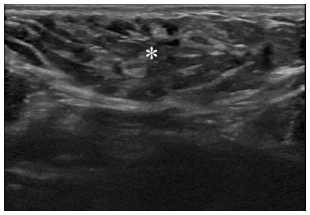
Figure 17. Figure 17. Accessory axillary breast tissue in a 36-year-old pregnant woman who presented with a right axillary lump for 2 months. US image shows heterogeneous echogenic tissue (*), similar to normal fibroglandular tissue, consistent with ac- cessory axillary breast tissue (BI-RADS 2).解説: 2ヶ月間右腋窩にしこりを訴えて来院した36歳の妊婦における、副乳の超音波画像を示します。US画像では、正常な線維腺組織と同様の不均一な高エコー組織(アスタリスク)が認められ、副乳と一致します(BI-RADS 2)。
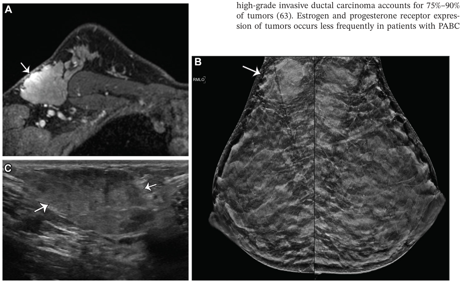
Figure 18. Figure 18. Incidental accessory axillary breast tissue in a 36-year-old lactating woman at high risk, with a family history of breast cancer. (A) Axial contrast-enhanced T1-weighted MR image shows an enhancing mass with irregular margins in the right axillary tail (arrow). Evaluation of the con- trast-enhancement kinetics showed rapid initial phase and delayed phase washout (not shown). (B) Bilateral mediolateral oblique mammograms show an irregular mass in the right axillary tail region (arrow). (C) Targeted US image of the right axilla shows an irregular heterogeneous mass with indis- tinct margins (arrows), which was categorized as BI-RADS 4A. US-guided biopsy of the mass confirmed benign breast tissue with lactational change.解説: 乳がんの家族歴を持つ高リスクの36歳の授乳中の女性における偶発的に発見された副乳の画像を示します。(A) 軸位造影T1強調MR画像は、右腋窩尾部に不規則な境界を持つ増強性腫瘤(矢印)を示しています。(B) 両側内外斜位マンモグラムは、右腋窩尾部領域に不規則な腫瘤(矢印)を示しています。(C) 右腋窩のターゲットUS画像は、不明瞭な境界を持つ不規則な不均一腫瘤(矢印)を示し、BI-RADS 4Aに分類されました。USガイド下生検で、授乳変化を伴う良性乳腺組織と診断されました。
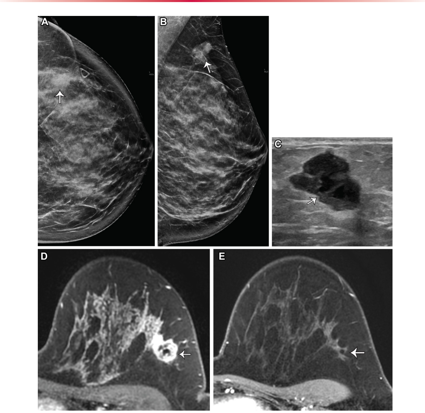
Figure 21. Figure 21. PABC in a 31-year-old woman who presented with a left breast lump. (A, B) Craniocaudal (A) and mediolateral oblique (B) mammograms show an irregular mass in the left breast, with indistinct margins (arrows), that corresponds with the overlying triangular marker in A. (C) US image shows a corresponding irregular hypoechoic mass with angular margins (ar- row) (BI-RADS 4C). US-guided biopsy results revealed grade 3 estrogen receptor–negative, progesterone receptor–negative, HER2–negative invasive ductal carcinoma. The patient subsequently discovered that she was pregnant but decided to termi- nate the pregnancy. MRI was performed after termination to evaluate for the extent of the disease. (D) Axial contrast-enhanced T1-weighted MR image shows the biopsy-proven malignant mass with a susceptibility artifact from the biopsy clip (arrow). No lymphadenopathy was seen. (E) Post–neoadjuvant chemotherapy contrast-enhanced T1-weighted MR image at the same level shows no residual mass or enhancement at the site of the biopsy-proven malignancy (arrow). The final pathologic results showed benign breast parenchyma with treatment effects and three negative sentinel lymph nodes.



コメントを残す