どうも、Beyond the Pixelです。肺の機能を評価する上で、放射線被曝のないMRI(磁気共鳴画像法)は非常に魅力的な画像診断装置です。しかし、そのMRI画像から肺の領域を正確に特定する「セグメンテーション」という作業は、CT(X線コンピュータ断層撮影)に比べて非常に難しいとされてきました。なぜなら、MRIは信号対雑音比(SNR)が低く、境界のコントラストが弱いため、自動セグメンテーションに必要な「アノテーション」(正確なラベル付け)を作成することが困難だからです。
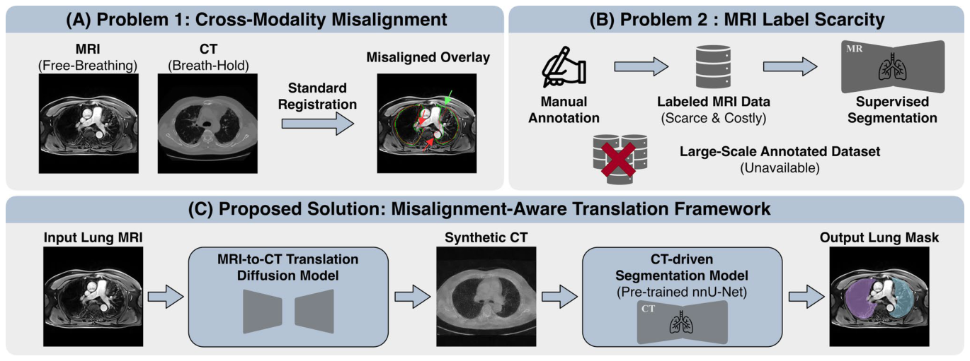
Figure 1. Fig. 1 Key challenges in lung MRI segmentation and the proposed solution. A Cross-modality misalignment: Differences in respira- tory states between free-breathing MRI and breath-hold CT lead to residual anatomical mismatches after standard registration, hindering direct label transfer. The lung contours derived from MRI (green) and CT (red) highlight the discrepancy, which is particularly pronounced解説: 肺MRIセグメンテーションにおける主要な課題と提案された解決策を示します。AはMRIとCT間のクロスモダリティ(異なる画像様式間)のズレを示し、呼吸状態の違いにより標準的なレジストレーション後も残存するズレがあることを指摘します。BはMRIラベルの希少性を示し、大規模で高品質なMRIアノテーションの入手が困難であることを説明します。Cは提案フレームワークの概要であり、NMI条件付き拡散モデルがラベルフリーのMRIから合成CTを生成し、標準的なCT訓練済みネットワークを使用して肺セグメンテーションを行うことを示しています。
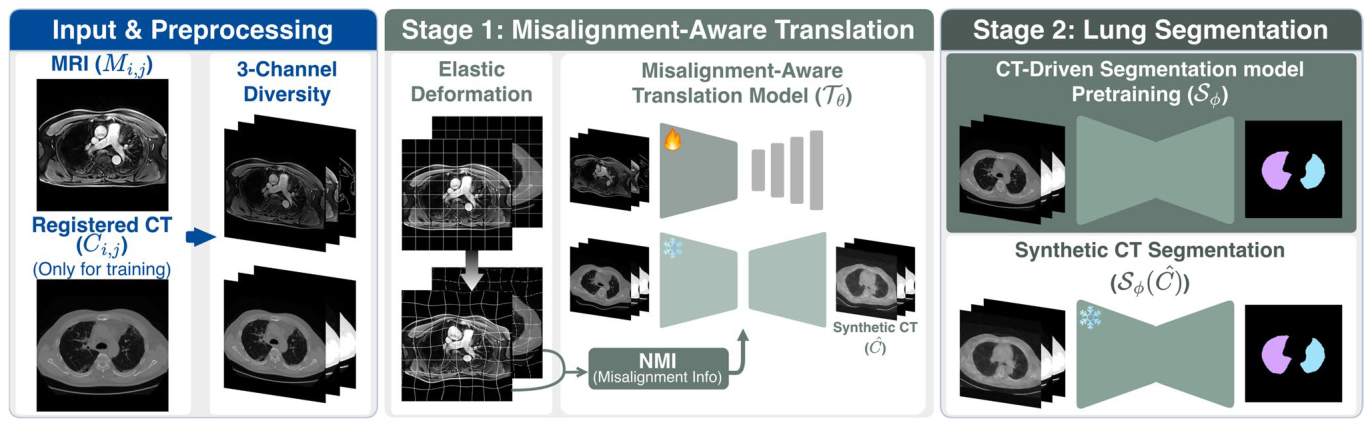
Figure 2. Fig. 2 Overview of the proposed framework. An unlabeled lung MRI is converted into synthetic CT (SynCT, ̂ C ) using a misalignment- aware diffusion model T휃 with three-channel diversity, elastic defor- mation, and NMI-based numerical conditioning. A CT-driven seg-解説: 提案されたフレームワークの全体像を示しています。入力されたラベルなし肺MRIは、3チャンネル多様性、弾性変形、およびNMIに基づく数値条件付けを備えたズレ対応型拡散モデルTθを用いて合成CT(SynCT, ̂C)に変換されます。その後、実際のCTで事前訓練されたCT駆動型セグメンテーションモデルSφがSynCTに適用され、肺マスクが取得されます。ペアのCTデータは、NMIの計算と翻訳モデルのガイドにのみ訓練中に使用されます。
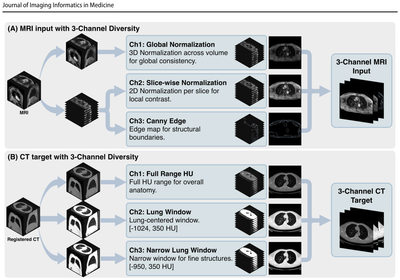
Figure 3. Fig. 3 Three-channel diversity design for MRI-to-CT translation. A For MRI, the three-channel input is formed by combining volume- level (3D) global normalization, slice-wise (2D) normalization, and a Canny edge map derived from the slice-wise normalized image to解説: MRIからCTへの翻訳のための3チャンネル多様性設計を示します。AはMRIの3チャンネル入力構成を示し、ボリュームレベル(3D)のグローバル正規化、スライスごと(2D)の正規化、スライスごとに正規化された画像から派生したCannyエッジマップを組み合わせて解剖学的境界を強調します。BはCTターゲットの3チャンネル構成を示し、全体的な解剖のためのフルレンジHU、標準的な肺ウィンドウ、および微細な肺実質構造を強調するための狭い肺ウィンドウの3つのHUウィンドウ設定を使用します。
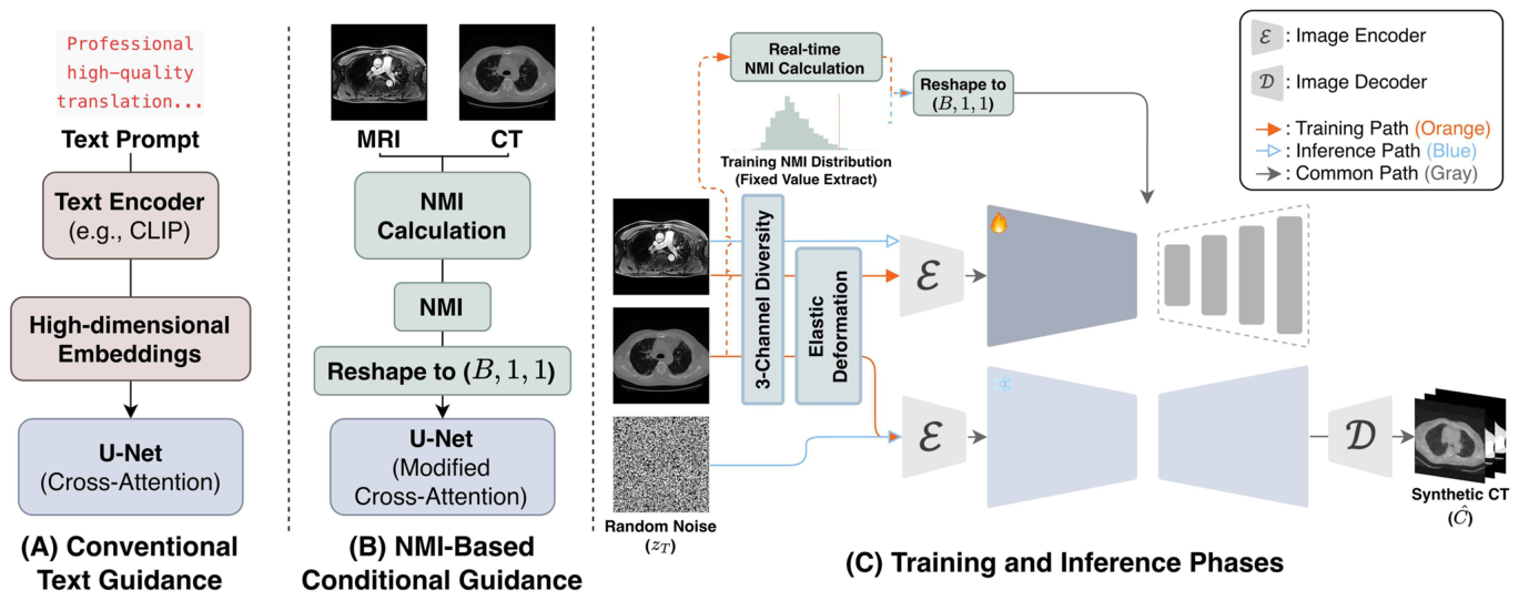
Figure 4. Fig. 4 NMI-based conditional guidance in diffusion translation. A Conventional diffusion models rely on text encoders and high-dimen- sional embeddings. B In the proposed framework, the text-condition- ing pathway is removed and replaced with scalar NMI-based condi- tioning, which is directly injected into the cross-attention blocks. C解説: 拡散翻訳におけるNMIに基づく条件付けのガイドを示します。Aはテキストエンコーダと高次元埋め込みに依存する従来の拡散モデルの条件付け方法を示します。Bは提案フレームワークにおけるNMIに基づくスカラー条件付けを示し、テキスト条件付け経路が削除され、NMIがクロスアテンションブロックに直接注入されます。Cは訓練および推論フェーズを示し、訓練中はペアのMRI-CTスライスからNMIが計算されて条件信号として使用され、推論中は訓練分布から導出された固定NMI値が適用されます。
研究チームは、Samsung Medical Centerで収集された、133ペアのMRI-CTスキャンを含む独自のデータセットを用いて実験を行いました。このデータセットには、訓練用に5537スライス、検証用に2605スライス、テスト用に2641スライスが含まれ、テストセットの27ペアのスキャンにのみMRIの肺セグメンテーションラベルが存在しました。訓練中はMRIアノテーションは一切使用されず、完全にラベルフリーの環境で評価されました。
基盤モデルの比較
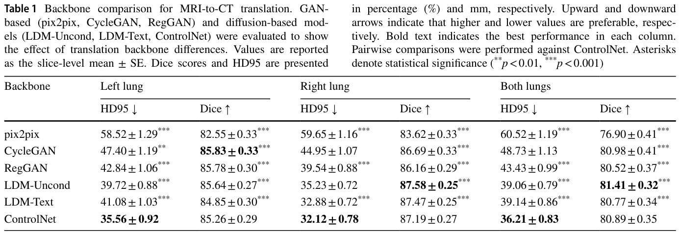
MRIからCTへの翻訳を行う様々な基盤モデルが比較されました。
Table 1. Table 1 Backbone comparison for MRI-to-CT translation. GAN- based (pix2pix, CycleGAN, RegGAN) and diffusion-based mod- els (LDM-Uncond, LDM-Text, ControlNet) were evaluated to show the effect of translation backbone differences. Values are reported as the slice-level mean ± SE. Dice scores and HD95 are presented解説: MRIからCTへの翻訳のための基盤モデル比較を示します。GANベース(pix2pix, CycleGAN, RegGAN)と拡散ベースモデル(LDM-Uncond, LDM-Text, ControlNet)が、翻訳基盤の違いの効果を示すために評価されました。ControlNetが最も安定した性能を示しました。
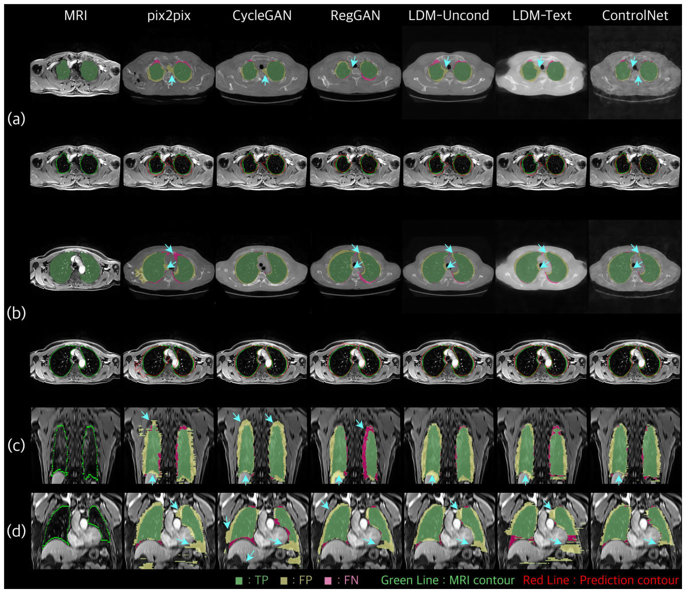
Figure 5. Fig. 5 Qualitative comparison of MRI-to-CT translation backbones. Four representative cases are shown in axial views (a, b) and coro- nal views (c, d). For (a, b), the top row shows error maps on SynCT (TP: green, FP: yellow, FN: magenta), and the bottom row overlays解説: MRIからCTへの翻訳基盤モデルの定性的な比較を示します。4つの代表的なケースが、軸方向ビュー(a, b)と冠状方向ビュー(c, d)で示されています。(a, b)では、上段はSynCT上のエラーマップ(TP:緑、FP:黄、FN:マゼンタ)を、下段はMRI上のグランドトゥルース(GT:緑)と予測された肺輪郭(予測:赤)を重ねて表示しています。(c, d)では、体積の一貫性を評価するためにMRI上の冠状エラーマップが提供されています。矢印は、基盤モデル間で顕著な境界の不一致がある領域を強調しています。
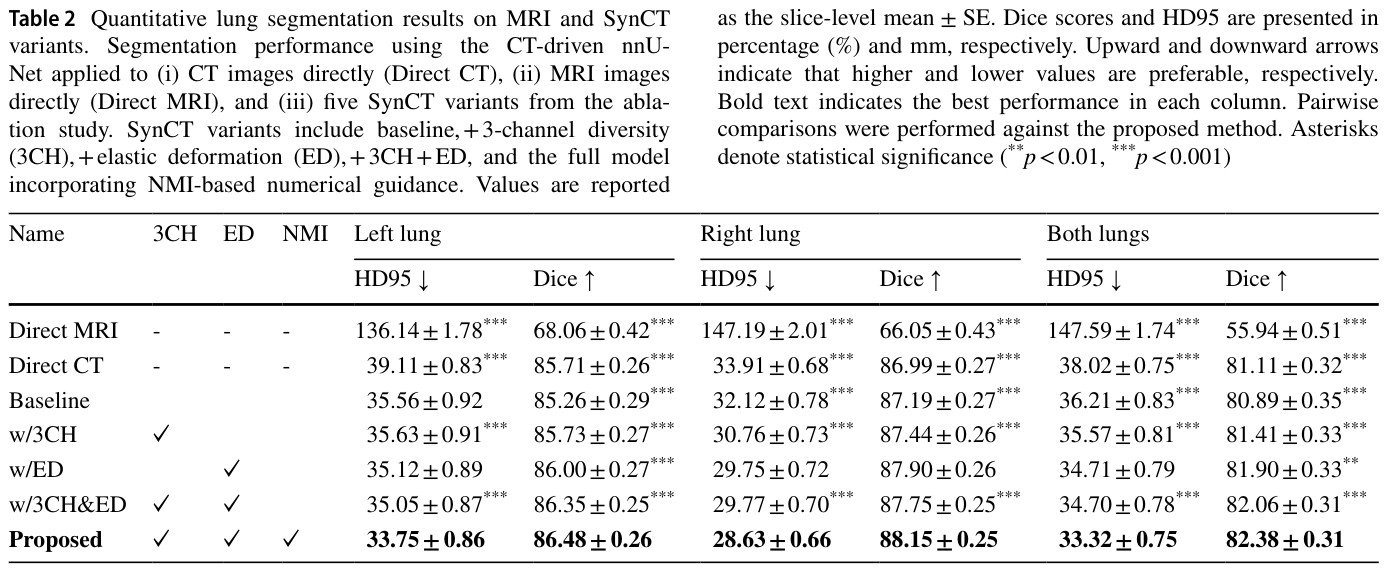
Table 2. Table 2 Quantitative lung segmentation results on MRI and SynCT variants. Segmentation performance using the CT-driven nnU- Net applied to (i) CT images directly (Direct CT), (ii) MRI images directly (Direct MRI), and (iii) five SynCT variants from the abla- tion study. SynCT variants include baseline, + 3-channel diversity (3CH), + elastic deformation (ED), + 3CH + ED, and the full model incorporating NMI-based numerical guidance. Values are reported解説: MRIおよびSynCTバリアントに関する定量的な肺セグメンテーション結果を示します。CT駆動型nnU-Netを(i)直接CT画像に、(ii)直接MRI画像に、(iii)アブレーション研究による5つのSynCTバリアントに適用した場合のセグメンテーション性能です。提案手法(Proposed)が両肺でDiceスコア82.38%、HD95 33.32 mmと最も良い性能を示しています。
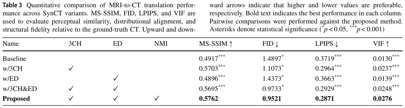
Table 3. Table 3 Quantitative comparison of MRI-to-CT translation perfor- mance across SynCT variants. MS-SSIM, FID, LPIPS, and VIF are used to evaluate perceptual similarity, distributional alignment, and structural fidelity relative to the ground-truth CT. Upward and down-解説: SynCTバリアントにおけるMRIからCTへの翻訳性能の定量比較を示します。MS-SSIM、FID、LPIPS、VIFを用いて、グランドトゥルースCTに対する知覚的類似性、分布アラインメント、構造的忠実度を評価しています。提案モデルが最も低いFIDスコアを達成し、最良の分布アラインメントを示しました。
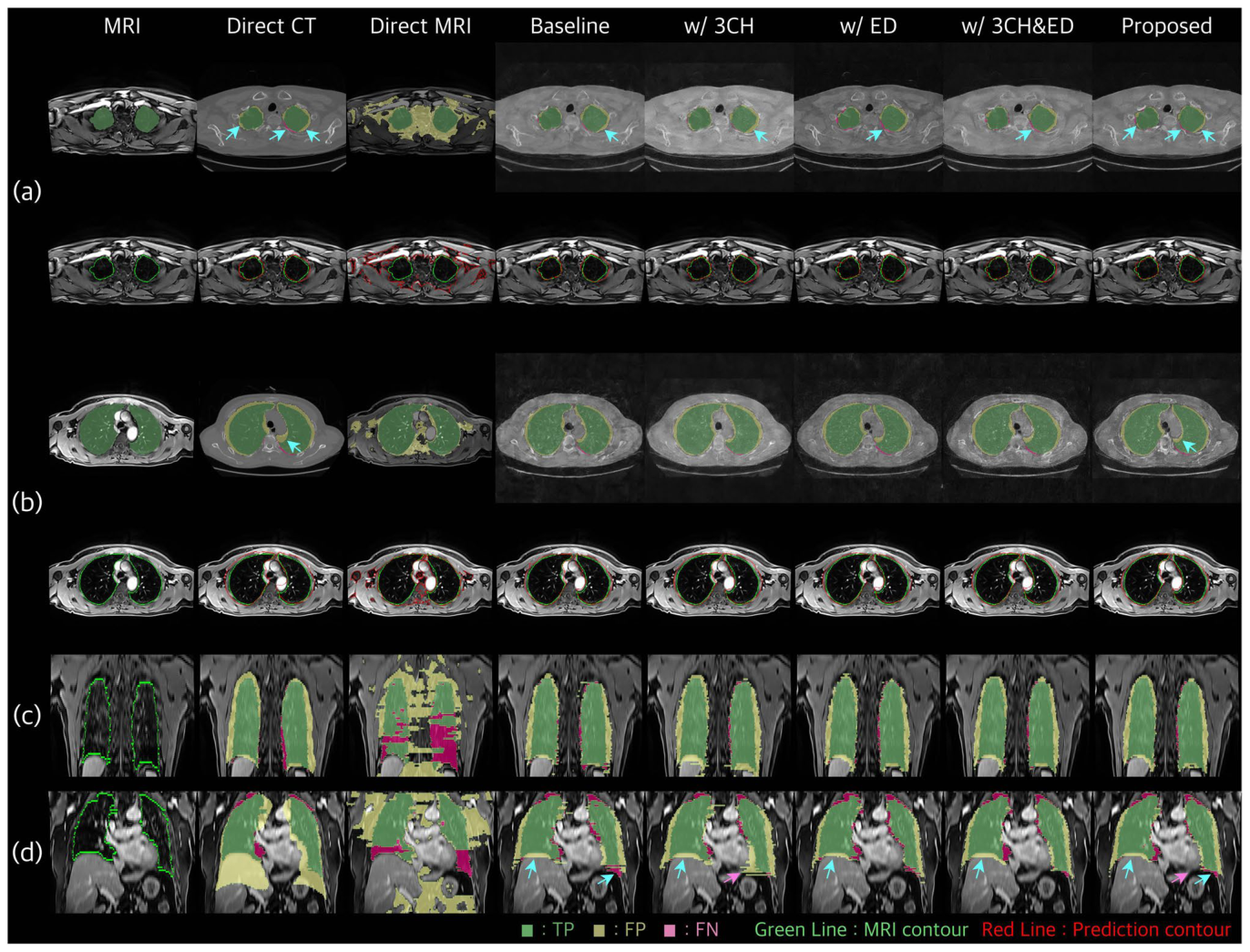
Figure 6. Fig. 6 Qualitative ablation results for the proposed framework. Four representative cases are shown in axial views (a, b) and coronal views (c, d). For (a, b), the top row shows error maps on SynCT (TP/FP/ FN), and the bottom row overlays ground-truth and predicted lung解説: 提案フレームワークの定性的なアブレーション結果を示します。4つの代表的なケースが軸方向ビュー(a, b)と冠状方向ビュー(c, d)で示されています。(a, b)では、上段はSynCT上のエラーマップ(TP/FP/FN)を、下段はMRI上のグランドトゥルースと予測された肺輪郭を重ねて表示しています。(c, d)では、体積の一貫性を示すためにMRI上の冠状エラーマップが提示されています。矢印は、個々の構成要素が境界の描写に影響を与える領域を示しています。
NMI条件付けの感度分析
NMI条件付けの値がセグメンテーション性能に与える影響も分析されました。
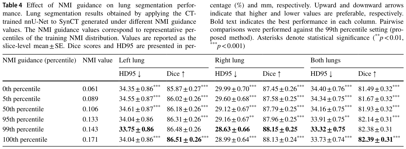
Table 4. Table 4 Effect of NMI guidance on lung segmentation perfor- mance. Lung segmentation results obtained by applying the CT- trained nnU-Net to SynCT generated under different NMI guidance values. The NMI guidance values correspond to representative per- centiles of the training NMI distribution. Values are reported as the slice-level mean ± SE. Dice scores and HD95 are presented in per-解説: NMIガイダンスが肺セグメンテーション性能に与える影響を示します。異なるNMIガイダンス値の下で生成されたSynCTにCT訓練済みnnU-Netを適用して得られた肺セグメンテーション結果です。99パーセンタイルが両肺でDiceスコア82.38%、HD95 33.32 mmと最も良い性能を示しています。
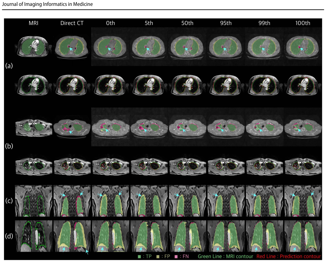
Figure 7. Fig. 7 Qualitative effect of NMI conditioning level on lung segmen- tation. Four representative cases are shown in axial views (a, b) and coronal views (c, d). For (a, b), the top row shows TP/FP/FN error maps on SynCT and the bottom row overlays ground-truth and pre- dicted lung contours on MRI across different NMI percentiles. For (c,解説: 肺セグメンテーションにおけるNMI条件付けレベルの定性的な効果を示します。4つの代表的なケースが軸方向ビュー(a, b)と冠状方向ビュー(c, d)で示されています。(a, b)では、上段はSynCT上のTP/FP/FNエラーマップを、下段は異なるNMIパーセンタイルでのMRI上のグランドトゥルースと予測された肺輪郭を重ねて表示しています。(c, d)では、様々なNMI条件付けレベルにおける体積の一貫性を評価するためにMRI上の冠状エラーマップが提供されています。NMI条件付けレベル間の違いは、アラインメント強度と構造的一貫性への影響を示しています。
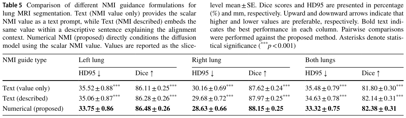
Table 5. Table 5 Comparison of different NMI guidance formulations for lung MRI segmentation. Text (NMI value only) provides the scalar NMI value as a text prompt, while Text (NMI described) embeds the same value within a descriptive sentence explaining the alignment context. Numerical NMI (proposed) directly conditions the diffusion model using the scalar NMI value. Values are reported as the slice-解説: 肺MRIセグメンテーションにおける異なるNMIガイダンス形式の比較を示します。テキスト(NMI値のみ)はスカラーNMI値をテキストプロンプトとして提供し、テキスト(NMI記述)は同じ値をアラインメントの文脈を説明する記述文に埋め込みます。数値NMI(提案手法)はスカラーNMI値を直接拡散モデルに条件付けします。数値NMI(提案手法)が両肺でDiceスコア82.38%、HD95 33.32 mmと最も良い性能を示しました。


コメントを残す