どうも、Beyond the Pixelです。心臓の血管である冠動脈が狭くなり、血流が滞ることで心筋に十分な酸素が供給されなくなる状態を「虚血性心疾患」と呼びます。この虚血の有無やその重症度を正確に診断することは、適切な治療方針を決定する上で非常に重要です。現在、血管ごとの虚血を評価する「ゴールドスタンダード」とされているのは、侵襲的冠動脈造影(ICA)というカテーテルを用いた検査中に測定される冠血流予備量比(FFR)や瞬間波形フリー比(iFR)です。これらは血管内の圧力差を測定することで血流障害の有無を評価しますが、体内にカテーテルを挿入する侵襲的な検査であるため、対象者への負担やコスト、合併症のリスクが伴います。
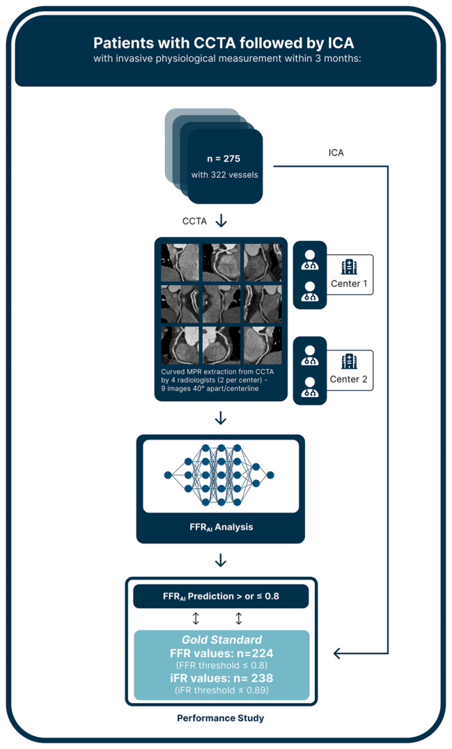
Figure 2. Fig. 2 Study workflow. All coronary computed tomography angiography (CCTA) examinations were evaluated by a senior and junior radiologist at each center for centerline-building supervision on all stenotic segments. A total of nine curved multiplanar reconstruction images of each coronary artery were uploaded to CorEx (Spimed-AI), a CCTA-based deep-learning model for invasive Fractional Flow Reserve prediction (CT-FFRAI). These images were subsequently analyzed to assess the diagnostic performance of CT-FFRAI, with invasive Fractional Flow Reserve ≤0.8 and invasive instantaneous wave-Free Ratio ≤0.89 as ground truth解説: 研究のワークフローを示しています。CCTA画像から専門家の監督のもと曲線状再構成画像(cMPR)が生成され、これをCT-FFRAI深層学習モデルが解析し、侵襲的FFR/iFR測定値をゴールドスタンダードとして診断性能が評価されるプロセスを表しています。
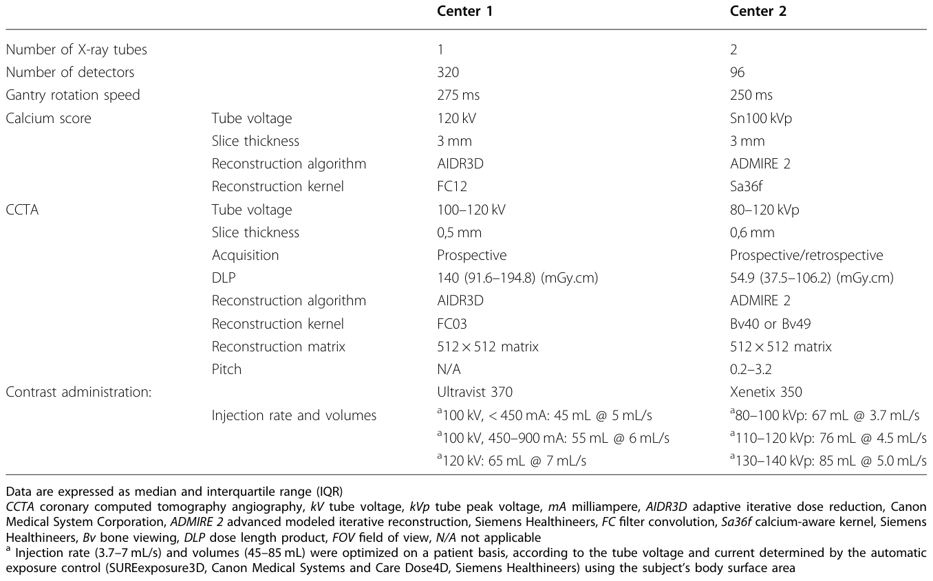
Table 1. Table 1 Technological and acquisition parameters for calcium score and CCTA imaging in both centers解説: 本研究に参加した2つの施設におけるカルシウムスコアおよびCCTA画像取得の技術的パラメータを示しています。X線管の数、検出器の数、ガントリー回転速度、スライス厚、造影剤注入条件などが施設間で異なることがわかります。
研究結果:AIの高い診断精度と石灰化の影響
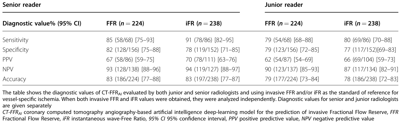
CT-FFRAIの二値アウトカム(0.80以下または0.80超)に関するシニア専門家とジュニア専門家による評価の一致度は「非常に良好(substantial)」と評価され(κ = 0.725, p < 0.001)、異なる経験レベルの専門家間でも一貫した結果が得られることが示されました。シニア専門家の監督下でのCT-FFRAIの診断精度は、侵襲的FFRが0.80以下の場合で83%(186/224)、侵襲的iFRが0.89以下の場合で83%(197/238)でした。
Table 3. Table 3 The diagnostic values of CT-FFRAI based on binary outcome analysis compared to invasive FFR and iFR measurements解説: シニアおよびジュニア専門家が評価したCT-FFRAIの診断値が示されており、侵襲的FFRおよびiFR測定値を参照標準としています。感度、特異度、陽性予測値、陰性予測値、診断精度がそれぞれパーセンテージと95%信頼区間で示されています。
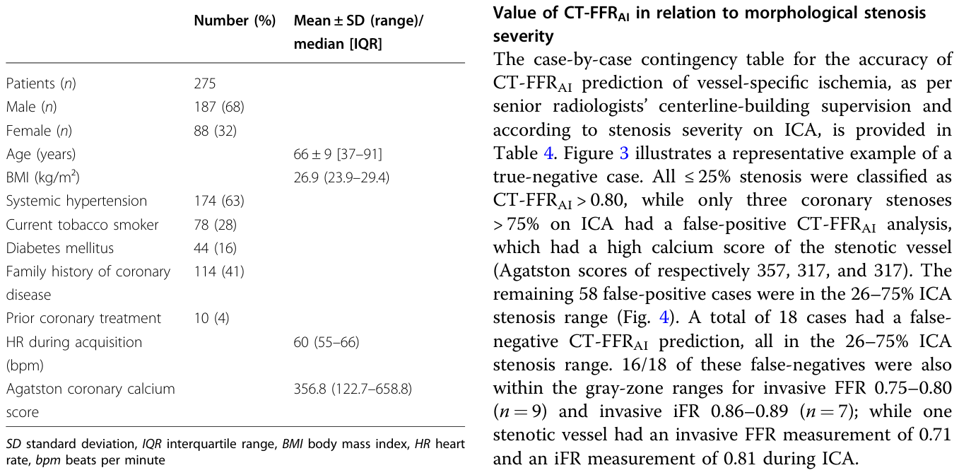
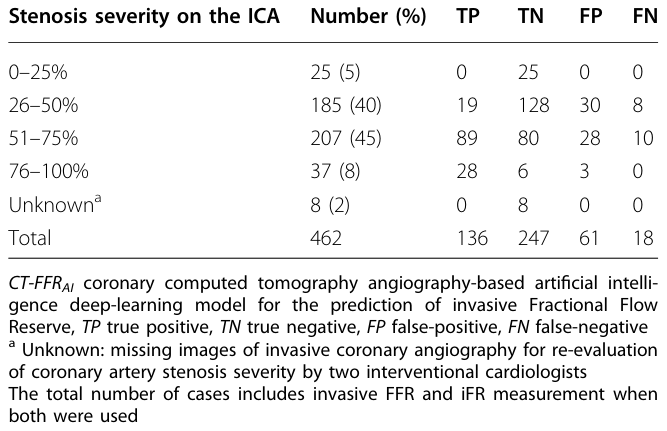
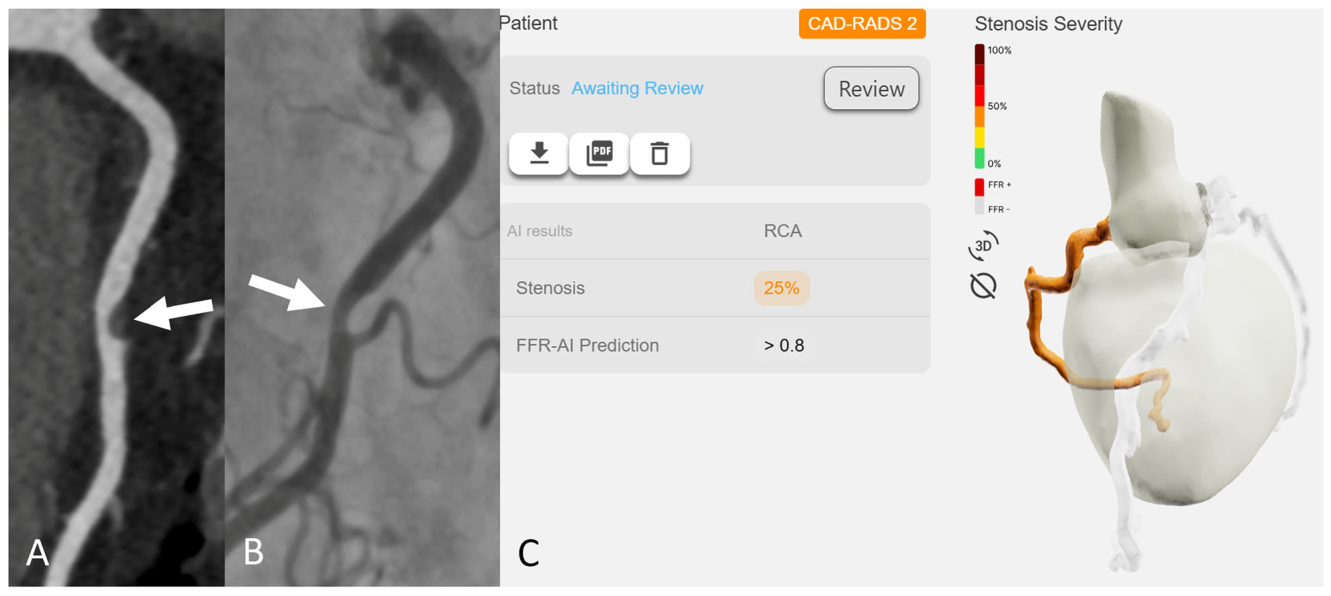
Table 4. Table 4 CT-FFRAI case-by-case assignment relative to vessel- specific ischemia based on senior radiologists' supervision and invasive coronary angiography stenosis severity解説: シニア専門家の監督に基づくCT-FFRAIの血管ごとの虚血予測と、侵襲的冠動脈造影(ICA)による狭窄度分類の関係を示す表です。狭窄度(0-25%から76-100%)ごとに真陽性、真陰性、偽陽性、偽陰性の数が示されています。Figure 3. Fig. 3 True-negative case. A representative case of a 60-year-old female patient with a recent onset of stable chest pain. Curved multiplanar reconstruction coronary computed tomography angiography A shows a soft plaque at the middle portion of the right coronary artery with obstructive stenosis (white arrow). Invasive coronary angiography B confirms a 26–50% stenosis (white arrow). The Fractional Flow Reserve measurement was 0.94, which is above the 0.8 threshold. Instantaneous wave-Free Ratio measurement was 0.95, which is above the 0.89 threshold. The CT-FFRAI prediction was > 0.8 (C)解説: 真陰性(虚血がないのにAIが虚血なしと判断し正しかった)ケースの代表例です。CCTA画像(A)と侵襲的冠動脈造影(B)で閉塞性狭窄が見られますが、侵襲的FFRとiFRの測定値は閾値より高く、CT-FFRAIも0.8超(虚血なし)と予測しています(C)。
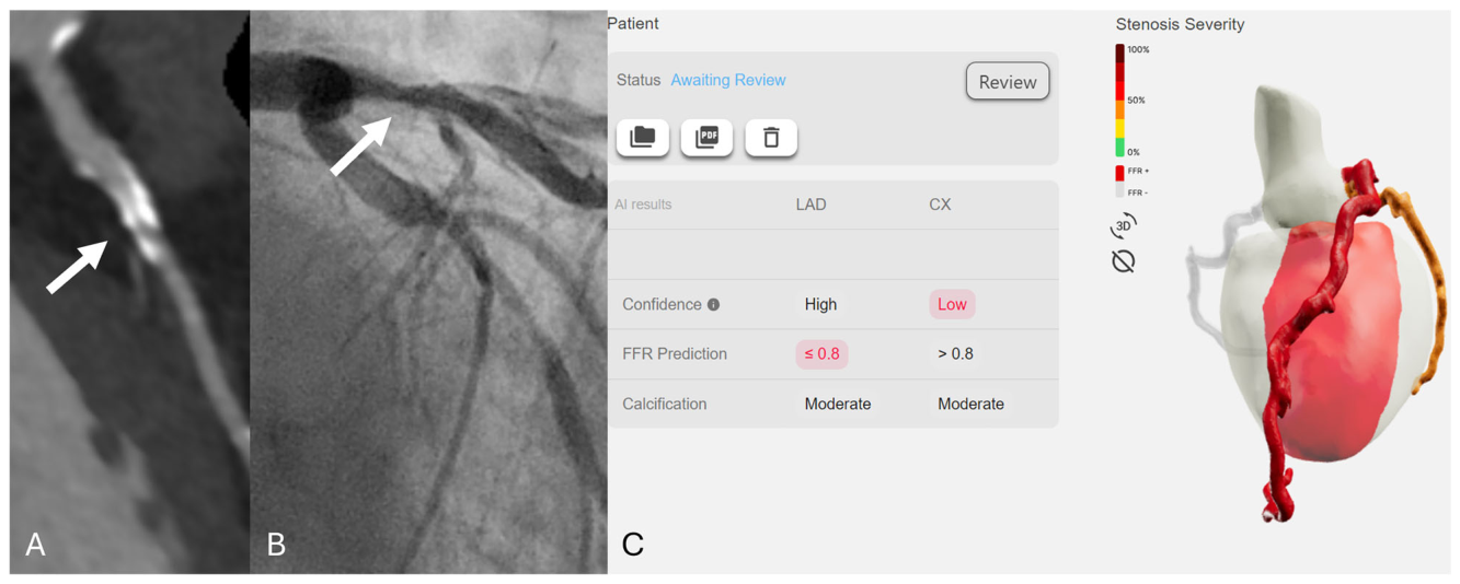
Figure 4. Fig. 4 False-positive case. A representative case of an 81-year-old male patient with a recent onset of stable chest pain. Curved multiplanar reconstruction coronary computed tomography angiography A shows a heavily calcified plaque with blooming artifacts (white arrow) in the proximal left anterior descending artery with obstructive stenosis. Invasive coronary angiography B demonstrates a 26–50% stenosis, and Fractional Flow Reserve was 0.89, in contrast to the report of CT-FFRAI that predicts FFR ≤0.8 (C). Of note, the patient also had an obstructive stenosis on the left circumflex artery with CT-FFRAI > 0.8 prediction and invasive Fractional Flow Reserve > 0.8 (FFR = 0.91)解説: 偽陽性(実際には虚血がないのにAIが虚血ありと判断してしまった)ケースの代表例です。CCTA画像(A)で高度石灰化を伴う閉塞性狭窄が見られます。侵襲的冠動脈造影(B)では狭窄が確認され、侵襲的FFRは閾値より高かったにもかかわらず、CT-FFRAIは0.8以下(虚血あり)と予測しています(C)。
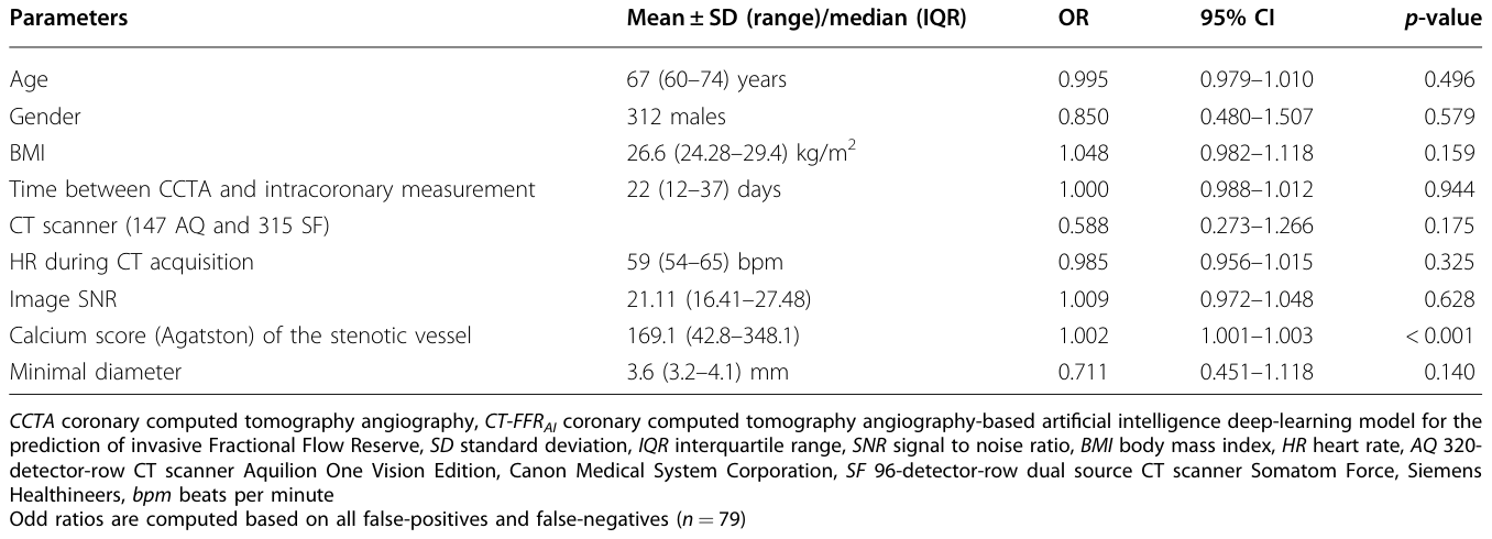
Table 5. Table 5 Multinomial logistic regression model with backward variable selection to identify patient- and CCTA image-related factors influencing the diagnostic performance of CT-FFRAI解説: CT-FFRAIの診断性能に影響を与える対象者およびCCTA画像関連因子を特定するための多項ロジスティック回帰モデルの結果です。年齢、性別、BMI、カルシウムスコアなどの因子と、そのオッズ比、95%信頼区間、p値が示されています。狭窄血管のカルシウムスコアが診断精度に有意な影響を与えたことがわかります。


コメントを残す