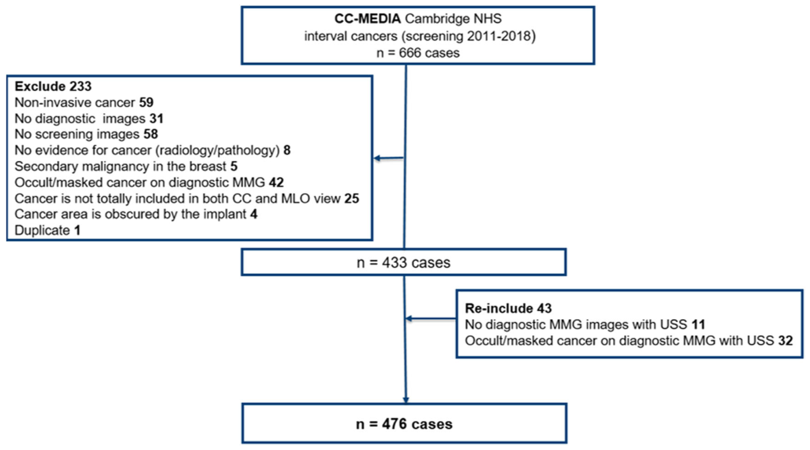
Figure 1. Fig. 1 A flow chart shows the number of initial interval cancer cases and the cases that were ruled out. CC, craniocaudal; MLO, mediolateral oblique; MMG, mammogram; NHS, National Health Service, USS, ultrasound解説: 本図は、研究に含める中間期がん症例の選択プロセスを示すフローチャートです。当初666例の中間期がん症例がありましたが、非侵襲性のがんや画像が利用できないケースなど233例が除外され、その後も一部のケースが再除外された結果、最終的に476例の中間期がんが解析対象となりました。
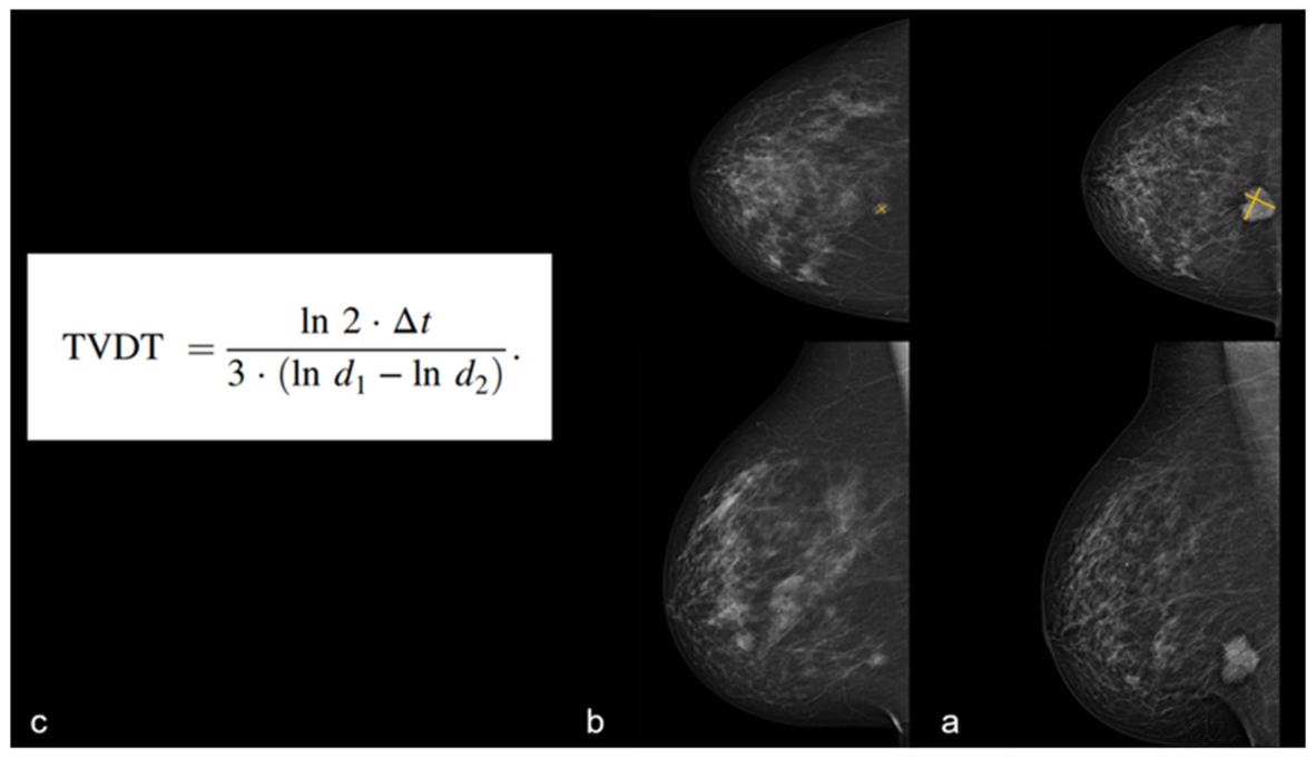
Figure 2. Fig. 2 Visible interval cancer at the time of screening: A 60-year-old female was diagnosed in the second year (931 days) following the screening with a 31 mm grade 3 ER-positive HER2 negative IDC with HGDCIS and LCIS. Using the same view (MLO view) on both diagnostic (a) and screening (b) MMG, mean size was measured based on the longest diameter and the true perpendicular diameter. Based on the mean size at both time points and the time interval in days (c), the calculated TVDT for this case was 230 days. HGDCIS, high-grade ductal carcinoma in situ; IDC, invasive ductal carcinoma; LCIS, lobular carcinoma in situ; MMG, mammogram; TVDT, tumour volume doubling time解説: 本図は、スクリーニング時に可視であった中間期がんの腫瘍体積倍加時間(TVDT)の計算式と例を示しています。左側にはTVDTの計算式があり、Δtはスクリーニングから診断までの日数、d1は診断時の平均直径、d2はスクリーニング時の平均直径です。右側には、診断時(a)とスクリーニング時(b)のマンモグラフィ画像が示され、がんは両時点で可視であり、そのサイズと診断までの期間に基づいてTVDTが230日と計算された症例が示されています。
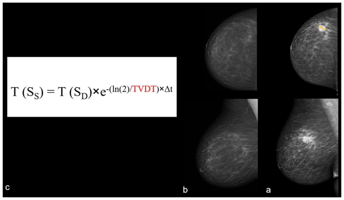
Figure 3. Fig. 3 Non-visible interval cancer at the time of screening: A 66-year-old female was diagnosed in the second year following the screening (758 days) with a 25 mm grade 3 ER-negative HER2 positive IDC with HGDCIS (a). Based on the patient’s age, cancer grade, and ER status, an estimated TVDT of 143 days was used in the equation (red, c). Together with the longest tumour diameter at diagnosis (SD) and time interval in days (Δt), a tumour size at screening (SS) was estimated at 0.7 mm (b). HGDCI, high-grade ductal carcinoma in situ; IDC, invasive ductal carcinoma; TVDT, tumour volume doubling time解説: 本図は、スクリーニング時に非可視であった中間期がんのサイズを推定する方法と例を示しています。左側にはスクリーニング時の腫瘍サイズ(SS)を推定するための式があり、診断時の腫瘍サイズ(SD)、推定TVDT、診断までの期間(Δt)を用いて計算されます。右側には、スクリーニングから758日後に診断された66歳の女性のマンモグラフィ画像が示されています(a, b)。この症例では、年齢、がんのグレード、ERステータスに基づき、TVDTの推定値が143日とされ(c)、スクリーニング時の腫瘍サイズは0.7mmと推定されました。
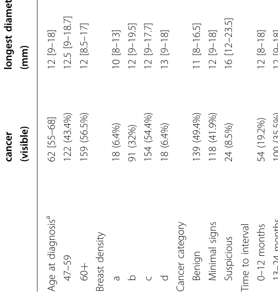
Table 2. Table 2 TVDT and size of the visible cancers at screening解説: 本表は、スクリーニング時に可視であった中間期がんについて、臨床病理学的変数(年齢、乳腺密度、がんの分類、診断までの期間、がんのグレード、ERステータス、HER2ステータス、分子サブタイプ)別に、スクリーニング時と診断時の最長径(mm)、診断までの日数、および腫瘍体積倍加時間(TVDT)の中央値と四分位範囲を示しています。TVDTは、年齢が高いほど長く、グレードが高いほど短く、ER陰性のがんで短いことが統計的に有意な差として示されています。
、
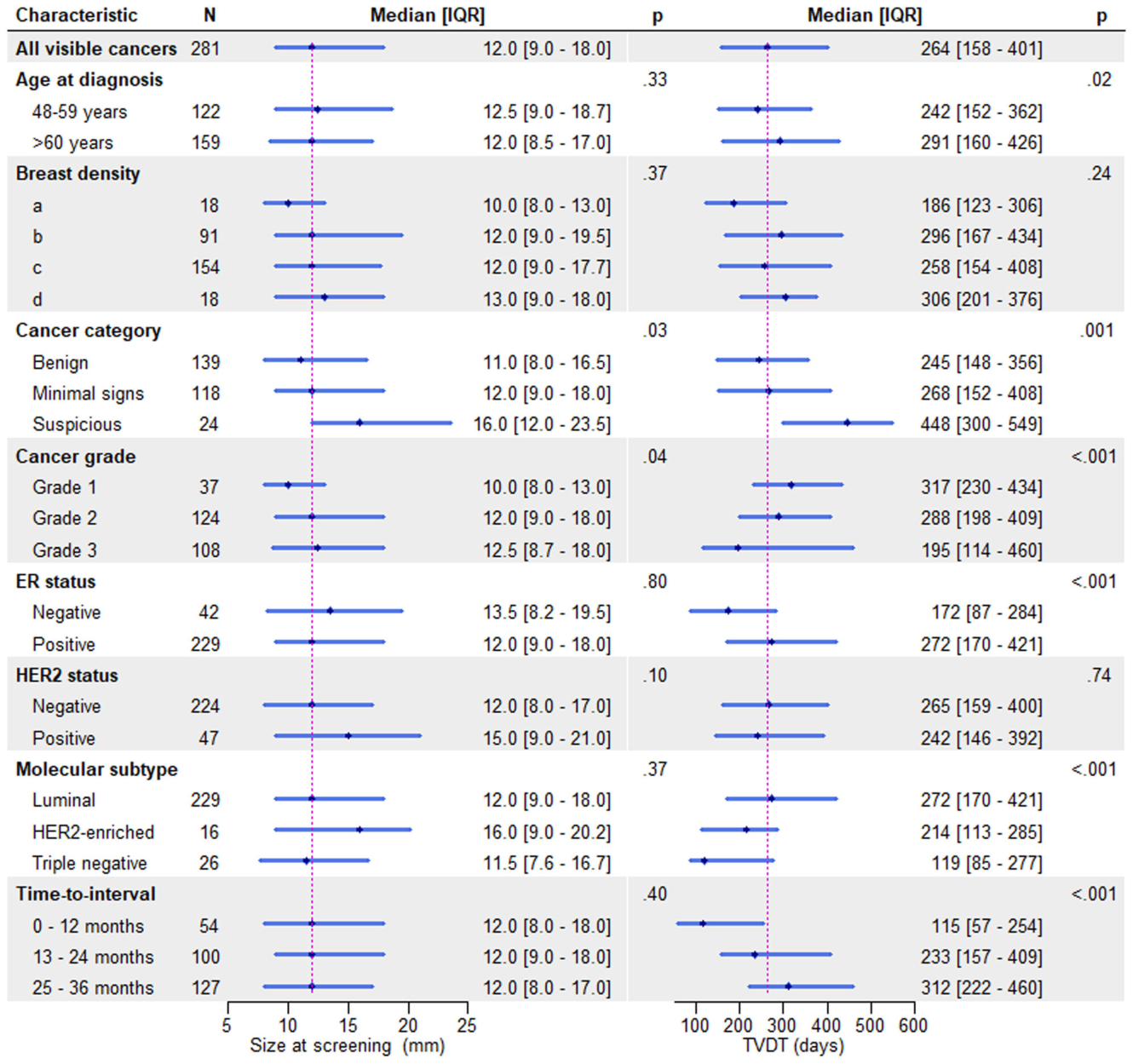
Figure 4. Fig. 4 Size at screening and tumour volume doubling time (TVDT) for visible interval cancers at screening (n = 281) stratified by clinicopathological characteristics: The magenta lines indicate the median size at screening and TVDT for all visible cancers. The dot and the length of each horizontal line denote the median and interquartile range (IQR), respectively. Size at screening represents the longest diameter解説: 本図は、スクリーニング時に可視であった中間期がん(n=281)について、がんの臨床病理学的特性別にスクリーニング時のサイズと腫瘍体積倍加時間(TVDT)の中央値と四分位範囲を示しています。TVDTは、年齢が高いほど長く、がんのグレードが高いほど短く、ER陰性のがんでは短い傾向が見られました。
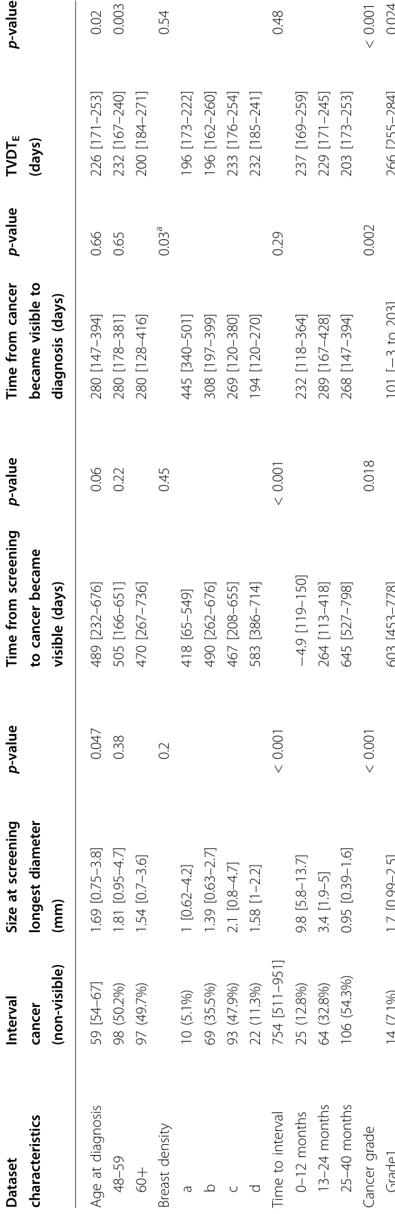
Table 3. Table 3 TVDT, cancer size estimation at screening, and cancer detectability time for non-visible cancers at 5 mm and 10 mm cancer size cut-offs for cancers developed in fatty解説: 本表は、スクリーニング時に非可視であった中間期がんについて、臨床病理学的変数別に、スクリーニング時の推定サイズ(mm)、がんが可視になるまでの推定時間(日)、可視から診断までの時間(日)、および推定腫瘍体積倍加時間(TVDTE)の中央値と四分位範囲を示しています。スクリーニングから診断までの期間が長いほど、スクリーニング時の推定サイズは小さい傾向にあります。また、グレード3やER陰性のがんではTVDTEが短いことが示されています。
、
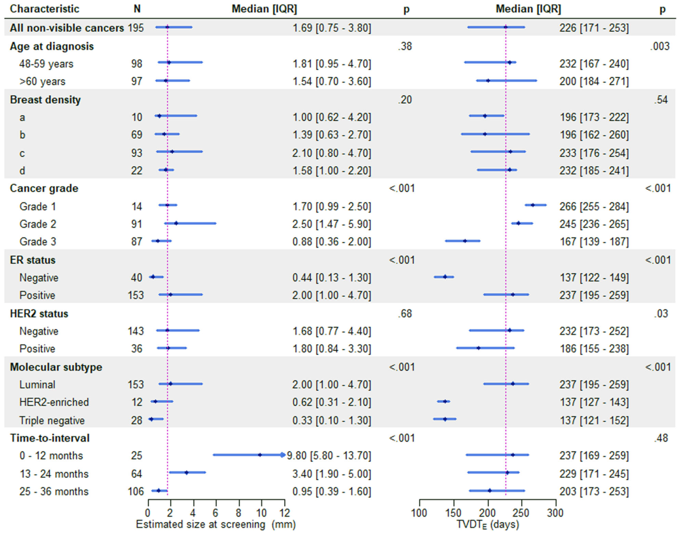
Figure 5. Fig. 5 Estimated tumour volume doubling time (TVDTE) and size at screening for non-visible interval cancers at screening (n = 281) stratified by clinicopathological characteristics: The magenta lines indicate the median size at screening and TVDT for all non-visible cancers. The dot and the length of each horizontal line denote the median and interquartile range (IQR), respectively. Size at screening represents the longest diameter解説: 本図は、スクリーニング時に非可視であった中間期がん(n=195)について、がんの臨床病理学的特性別にスクリーニング時の推定サイズと推定腫瘍体積倍加時間(TVDTE)の中央値と四分位範囲を示しています。TVDTEは、年齢が若いほど長く、がんのグレードが高いほど短く、ER陰性のがんでは短い傾向が見られました。


コメントを残す