どうも、Beyond the Pixelです。がん治療における分子メカニズムの理解が進むにつれて、「精密がん治療」という新しい時代が到来しました。この治療法は、がんの発生や進行に関わる特定の分子を標的とし、多くの対象者に顕著な治療効果をもたらしています。しかし、その一方で、特定の薬剤に特有のさまざまな合併症を伴うことも明らかになっています。特に、免疫チェックポイント阻害薬の使用が急速に拡大する中で、「免疫関連有害事象」と呼ばれる特有の毒性反応の診断とモニタリングが新たな課題となっています。がんのケアにおいて重要な役割を果たす放射線専門家にとって、最先端のがん治療アプローチに精通し、新しい治療法によって生じる合併症を認識することは不可欠です。本稿は、精密がん治療における胸部合併症について包括的にレビューし、その画像所見と臨床的特徴を説明し、これらの病変の診断とモニタリングにおける放射線専門家の役割を議論します。また、胸部合併症に関連する抗がん剤の分子メカニズムにも言及し、新規がん治療における新たな課題を強調しています。この記事は、精密がん医療の時代における放射線専門家の日常診療のための実用的な参考ガイドとして役立つように設計されています。
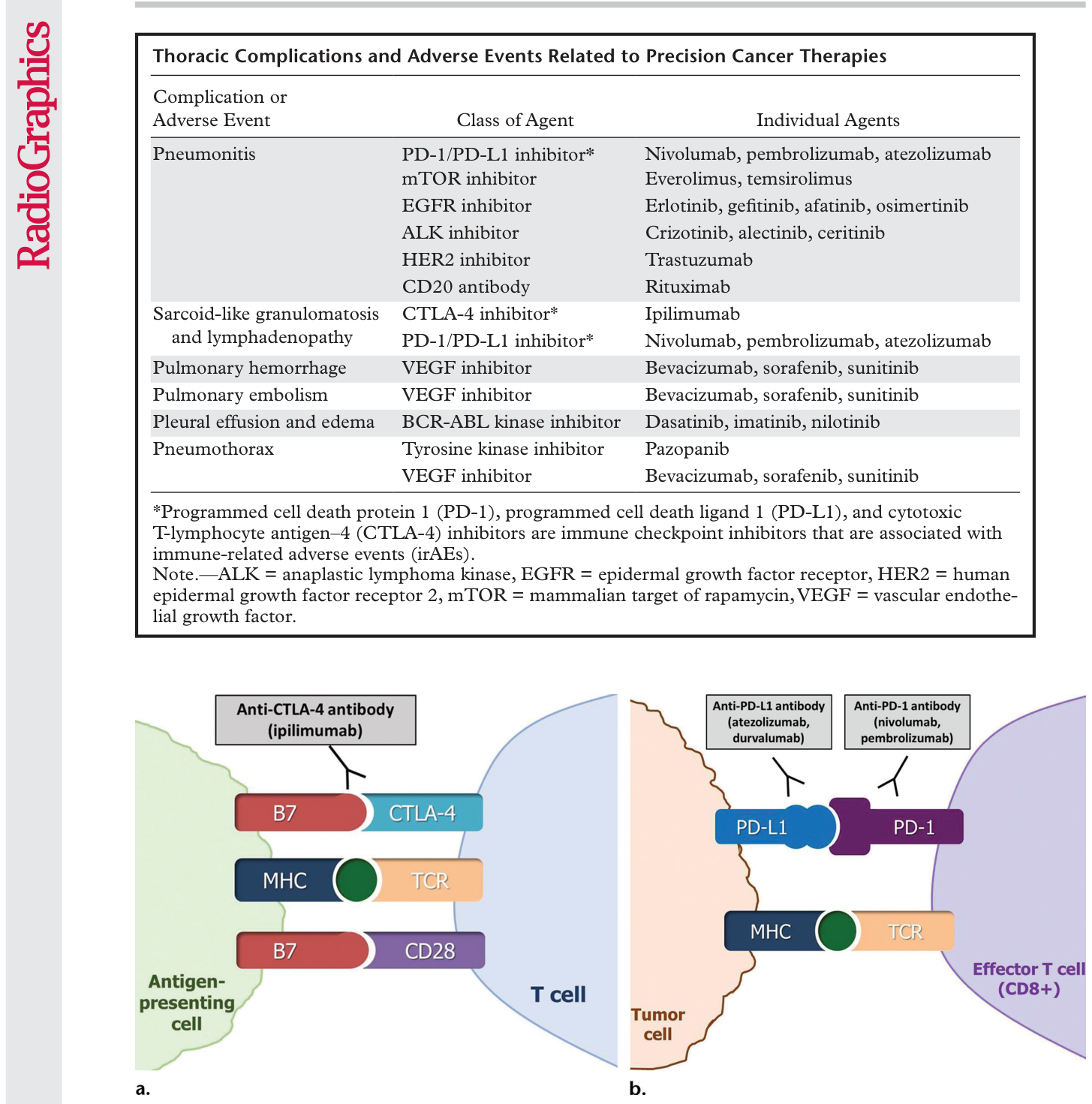
Figure 1. Figure 1. Diagram of immune checkpoint inhibitor therapy shows immune inhibition by tumors and its blockade as the mecha nism of action. MHC = major histocompatibility complex, TCR = T-cell receptor. (a) CTLA-4 is an immune checkpoint molecule on T cells, and its interaction with the ligand B7 on antigen-presenting cells causes inhibition of the T-cell immune response against the tumor, which allows the tumor cells to evade immune attack. CTLA-4 inhibitors such as ipilimumab prevent this interaction by binding to CTLA-4 on T cells and blocking T-cell immune inhibition, thereby activating an immune response against tumor cells. (b) The binding of PD-1 on effector T cells and of its ligand PD-L1 on tumor cells delivers an inhibitory signal that decreases cytokine production and T‑cell proliferation, which allows tumor cells to escape from immune response. PD-1 or PD-L1 inhibitors block the binding and prevent tumoral immune inhibition, thus inducing an antitumor immune response. PD-1/PD-L1 blockade by agents such as nivolumab, pembrolizumab, atezolizumab, and durvalumab has become a major treatment option for many types of advanced cancers. (Adapted and reprinted under a CC BY 4.0 license from reference 8.)解説: 免疫チェックポイント阻害薬がどのようにT細胞の活性化を促し、腫瘍に対する免疫応答を活性化するかを示す図です。CTLA-4阻害薬やPD-1/PD-L1阻害薬が、腫瘍が免疫系から逃れるのを防ぐメカニズムが示されています。
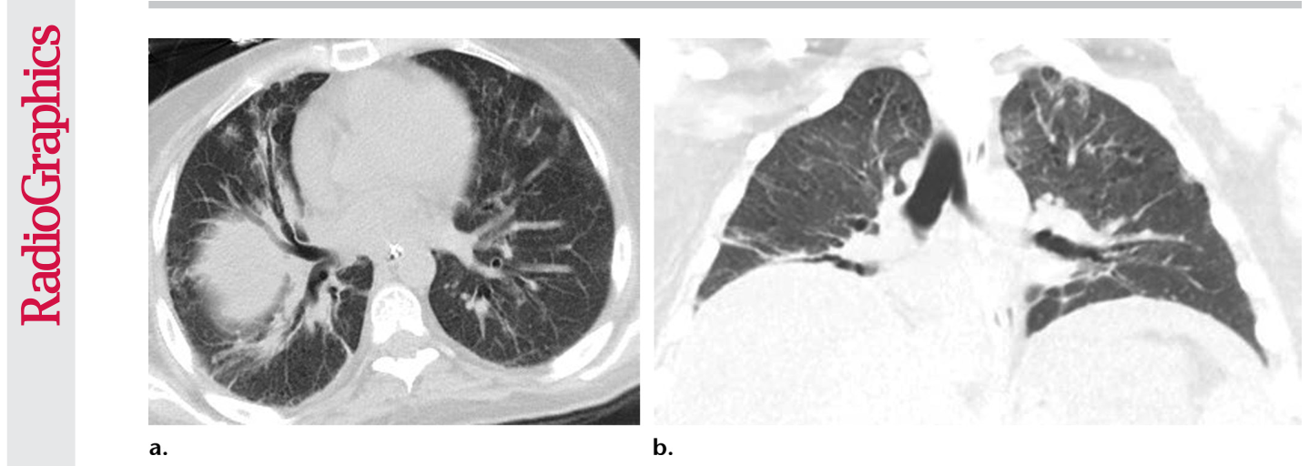
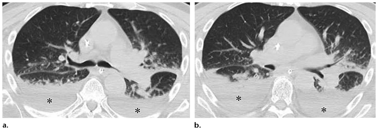
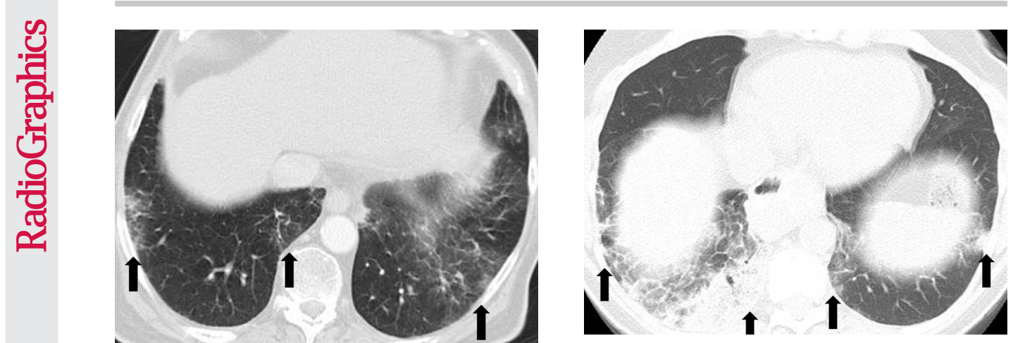
Figure 2. Figure 2. PD-1 inhibitor pneumonitis: AIP/ARDS pattern in a 38-year-old woman with advanced melanoma who was treated with nivolumab. Axial (a) and coronal (b) chest CT images obtained at 15 weeks of therapy show diffuse GGOs and traction bronchiec tasis, with markedly decreased lung volumes seen on the coronal image and an elevated right hemidiaphragm, findings indicative of a radiologic AIP/ARDS pattern. The patient was admitted to the intensive care unit, was treated with intravenous corticosteroids, and also required infliximab (an anti–tumor necrosis factor-α immunosuppressive agent) therapy.解説: 進行メラノーマの38歳女性においてニボルマブ治療15週後に撮影された胸部CT画像です。びまん性のすりガラス陰影と牽引性気管支拡張、著しい肺容積の減少、右横隔膜の挙上が見られ、放射線学的なAIP/ARDSパターンを示しています。Figure 3. Figure 3. Pneumonitis with AIP/ARDS pattern in a 70-year-old man with melanoma who was treated with sequentially administered nivolumab and ipilimumab combination therapy. Axial chest CT images obtained at 5.6 months of therapy (a at the level of the carina; b at a lower level than a) show GGOs, reticular opacities, consolidation, and traction bronchiectasis, as well as pleural effusions (*) involving both lungs. (Figure reprinted from reference 7.)解説: メラノーマの70歳男性がニボルマブとイピリムマブの併用療法を受けて5.6ヶ月後に撮影された胸部CT画像です。両肺にすりガラス陰影、網状影、浸潤影、牽引性気管支拡張、そして胸水(*印)が見られ、AIP/ARDSパターンの肺臓炎を示しています。
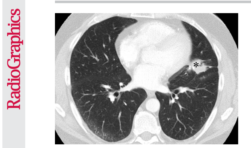
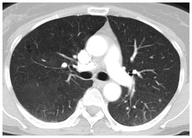
Figure 4. Figure 4. PD-1 inhibitor pneumonitis: NSIP pattern in a 58-year-old man with advanced melanoma who was treated with nivolumab. Axial chest CT image obtained at 7 weeks of therapy shows GGOs and reticular opacities in a subpleural distribution, representing an NSIP pattern of PD-1 inhibitor pneumonitis. * = metastatic lesion in the lung.解説: 進行メラノーマの58歳男性がニボルマブ治療7週後に撮影された胸部CT画像です。胸膜下にすりガラス陰影と網状影が見られ、PD-1阻害薬による肺臓炎のNSIPパターンを示しています。(*印は肺転移病変)。
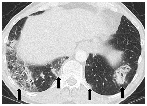
Figure 5. Figure 5. PD-1 inhibitor pneumonitis: COP pattern in a 69-year-old man with advanced NSCLC who was treated with nivolumab. At 6 months of therapy, the patient presented with increased shortness of breath and cough, without fever. Axial chest CT image shows multifocal areas of consolidation and GGOs in a predominantly peripheral and basilar distribution (arrows), representing a COP pattern of PD-1 inhibitor–related pneumonitis. Bronchial dilatation was noted within the areas of consolidation. Nivolumab was withheld, and the patient was treated with corticosteroids, with subsequent improvement.解説: 進行非小細胞肺がんの69歳男性がニボルマブ治療6ヶ月後に撮影された胸部CT画像です。主に末梢および肺底部に多発性の浸潤影とすりガラス陰影(矢印)が見られ、PD-1阻害薬関連肺臓炎のCOPパターンを示しています。浸潤影内には気管支拡張が認められます。
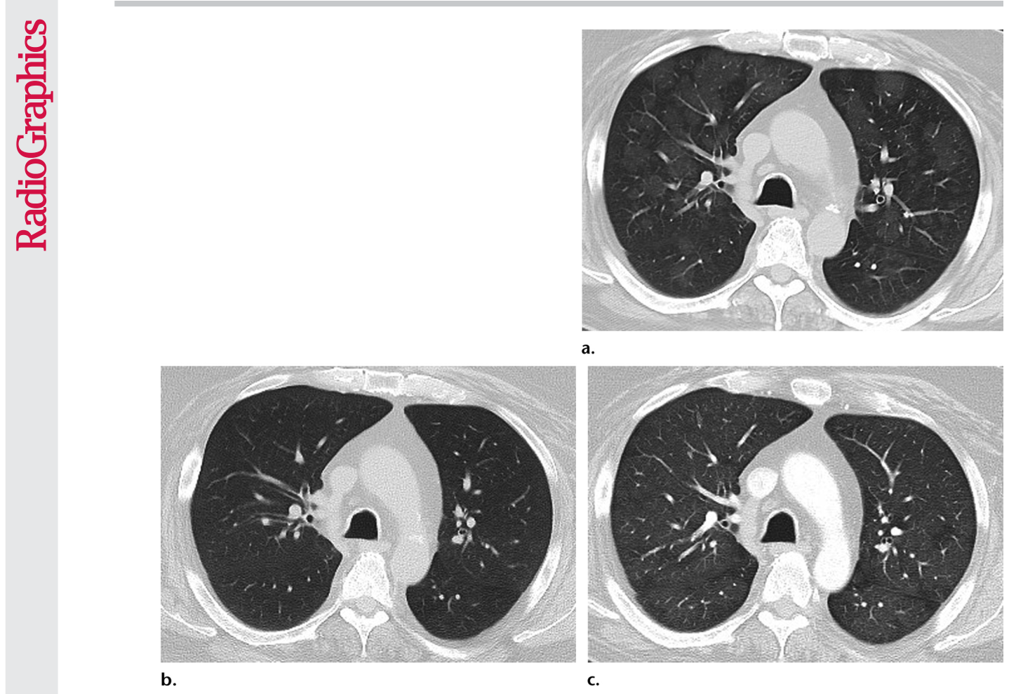
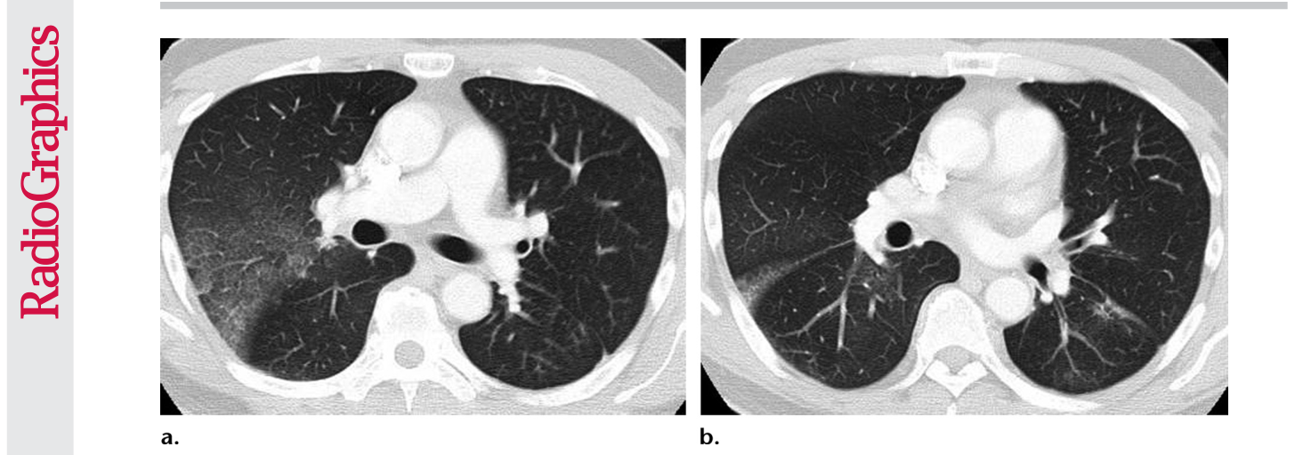
Figure 6. Figure 6. PD-1 inhibitor pneumonitis: Hypersensitivity pneumonitis pattern in a 68-year-old man with metastatic renal cell carcinoma who was treated with nivolumab and presented with a new cough at 6 months of therapy. (a) Axial chest CT im age shows new multifocal GGOs in a centrilobular distribution throughout both lungs and mosaic attenuation, findings that represent pneumonitis with a hypersensitivity pneumonitis pattern. Nivolumab therapy was withheld, and the patient under went corticosteroid therapy. (b) Axial follow-up CT image obtained after 1 month of corticosteroid therapy shows marked improvement of pneumonitis and resolution of GGOs. Nivolumab continued to be withheld, and corticosteroid therapy was tapered. At 1.5 months after completing the corticosteroid taper, without restarting nivolumab or any other systemic therapy, the patient experienced a worsening cough. (c) Axial chest CT image shows development of diffuse GGOs with areas of air trapping, findings indicative of pneumonitis with a hypersensitivy pneumonitis pattern. The radiographic pattern of this sec ond episode is similar to that noted in the initial episode and represents pneumonitis flare.解説: 転移性腎細胞がんの68歳男性がニボルマブ治療6ヶ月後に現れた、過敏性肺炎パターンのPD-1阻害薬肺臓炎を示す胸部CT画像です。(a) 小葉中心性に多発性のすりガラス陰影とモザイク状の濃度変化が見られます。(b) コルチコステロイド治療1ヶ月後の改善。(c) コルチコステロイド治療完了後、ニボルマブ非再開での肺臓炎フレアを示すびまん性すりガラス陰影とエアトラッピングです。
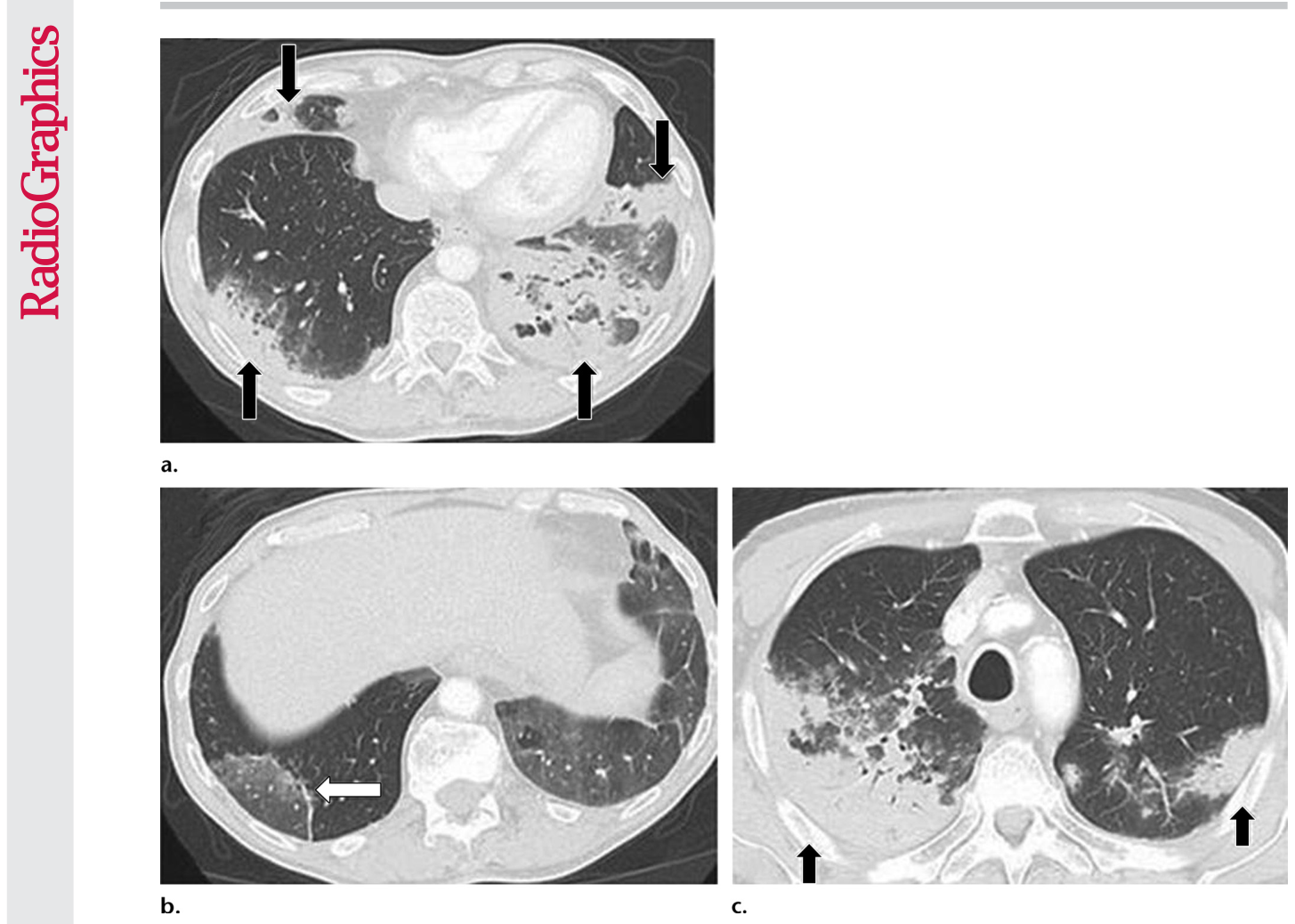
Figure 7. Figure 7. PD-1 inhibitor pneumonitis flare in a 72-year-old man with stage IV squamous NSCLC who was treated with nivolumab and presented with progressive dyspnea with cough and wheezing but no fever. (a) Axial chest CT image at 15 weeks of therapy demonstrates multifocal areas of GGOs, reticular opacities, and consolidation (arrows) involving all lobes, as well as centrilobular nodularity and traction bronchiectasis in a predominantly peripheral distribution. The overall features demonstrate a COP pattern. The patient was treated with prednisone for pneumonitis. (b) Axial follow-up CT image after 4 weeks of prednisone therapy shows a significant decrease in the findings, with residual GGOs. Note the “reversed halo” sign manifesting as a central GGO surrounded by a crecent-shaped dense airspace consolidation (arrow), a finding that has been reported as a radiologic manifestation of COP. (c) Axial CT image obtained 4 weeks after the completion of prednisone therapy shows the development of a bilateral dense consolidation with GGOs and reticular opacities (arrows) in peripheral and multifocal distributions, again demonstrating a COP pattern as noted during the first episode of PD-1 inhibitor pneumonitis. Given the similarity of the radiologic and clinical manifesta tions to those of the first episode, the patient restarted prednisone for treatment of pneumonitis flare. Follow-up chest CT images obtained 2 weeks after starting the second course of prednisone therapy (not shown) demonstrated a decrease in the findings, indicative of improving pneumonitis in response to corticosteroid therapy. (Figure 7 reprinted from reference 21.)解説: ステージIVの扁平上皮非小細胞肺がんの72歳男性がニボルマブ治療15週後に発症したPD-1阻害薬肺臓炎フレアの胸部CT画像です。(a) 多発性のすりガラス陰影、網状影、浸潤影(矢印)を示し、COPパターンを示しています。(b) プレドニゾン治療4週後の所見の著しい減少。(c) プレドニゾン治療完了4週後に再発したCOPパターンの両側性浸潤影とすりガラス陰影(矢印)です。
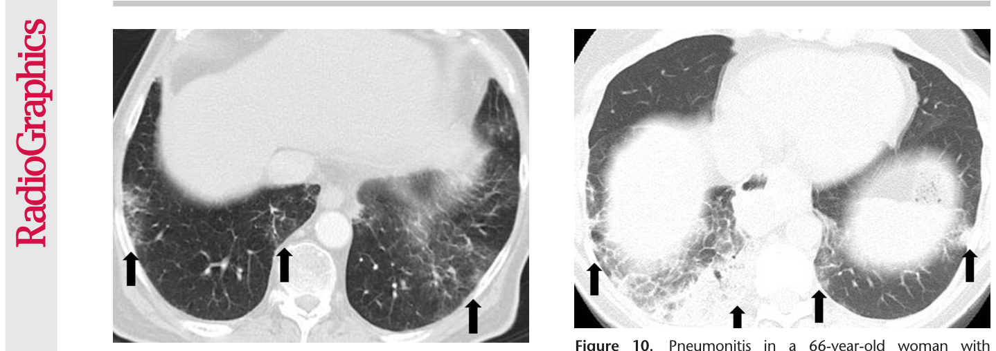
Figure 9. Figure 9. Pneumonitis in a 71-year-old man with metastatic renal cell carcinoma treated with temsirolimus. Axial CT im age at 4 weeks of therapy shows multifocal GGOs and reticular opacities in a predominantly peripheral and basilar distribution (arrows), findings that represent mTOR inhibitor–related pneu monitis. The patient was symptomatic and was switched to an alternate therapy.解説: 転移性腎細胞がんの71歳男性がテムシロリムス治療4週後に撮影されたCT画像です。主に末梢および肺底部に多発性のすりガラス陰影と網状影(矢印)が見られ、mTOR阻害薬関連肺臓炎を示しています。Figure 10. Figure 10. Pneumonitis in a 66-year-old woman with Waldenström macroglobulinemia treated with mTOR inhibitor therapy. Axial CT image at 6 months of therapy shows consoli dation, GGOs, and reticular opacities (arrows) that represent a COP pattern.解説: ワルデンシュトレームマクログロブリン血症の66歳女性がmTOR阻害薬治療6ヶ月後に撮影されたCT画像です。浸潤影、すりガラス陰影、網状影(矢印)が見られ、COPパターンを示しています。
CT所見には、すりガラス陰影(GGOs)、浸潤影、網状影が含まれます。
Figure 11. Figure 11. Pneumonitis in a 62-year-old woman with ad vanced pancreatic neuroendocrine tumor treated with evero limus and temozolomide. Axial CT image obtained at 10.3 months of therapy shows diffuse bilateral GGOs and reticular opacities that are indicative of a hypersensitivity pneumonitis pattern. The patient had mild shortness of breath and was treated with prednisone.解説: 進行膵神経内分泌腫瘍の62歳女性がエベロリムスとテモゾロミド治療10.3ヶ月後に撮影されたCT画像です。びまん性の両側性すりガラス陰影と網状影が見られ、過敏性肺炎パターンを示しています。
Figure 12. Figure 12. Pneumonitis in a 42-year-old man with an EGFR exon 19 deletion mutation who was treated with erlotinib in the United States. Axial chest CT images (b obtained at a lower level than a) obtained at 8 weeks of therapy show multifocal areas of GGOs in both lungs, findings that represent pneumonitis. Note the absence of traction bronchietasis or volume loss.解説: 米国でエルロチニブ治療を受けたEGFRエクソン19欠失変異を持つ42歳男性の胸部CT画像です。治療8週後に両肺に多発性のすりガラス陰影が見られ、肺臓炎を示しています。牽引性気管支拡張や肺容積の減少は見られません。
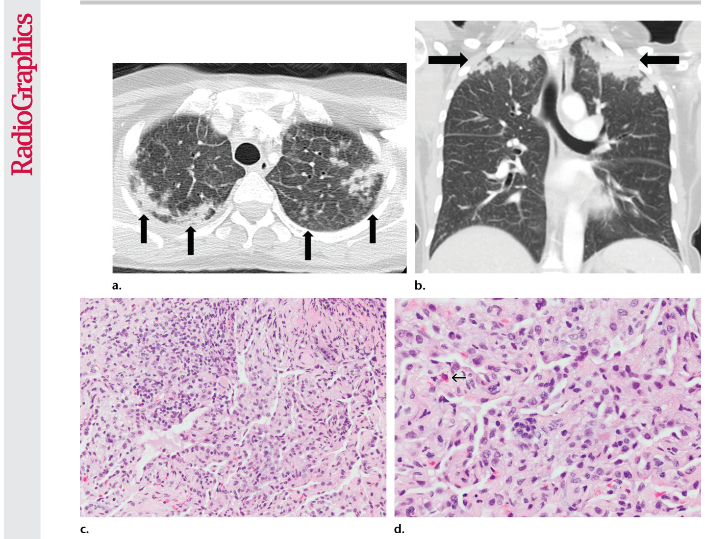
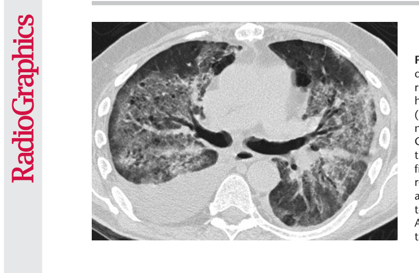
Figure 13. Figure 13. ALK inhibitor–related pneumonitis in a 55-year-old woman with ALK-positive stage IV adenocarcinoma of the lung. The patient experienced disease progression while taking a first-generation ALK inhibitor (crizotinib) and was then treated with a sec ond-generation ALK inhibitor (ceritinib). She presented with an increasing dry cough and dyspnea at 7 months of ceritinib therapy. (a, b) Axial (a) and coronal (b) CT images show biapical consolidation and GGOs in both upper lobes (arrows) in a striking peripheral distribution, indicative of a COP pattern. (c, d) Photomicrographs from transbronchial lung biopsy specimen show organizing interstitial pneumonia characterized by alveolar interstitial widening by lymphocytic infiltrates, increased extracellular matrix material, reactive pneumocyte hyperplasia, scattered eosinophils (arrow in d), and numerous airspace foamy macrophages. There was no evidence of tumor in the biopsy specimen. (Hematoxylin-eosin stain; original magnification, 3200 in c, 3400 in d.)解説: ALK陽性ステージIV肺腺がんの55歳女性におけるALK阻害薬関連肺臓炎です。セリチニブ治療7ヶ月後に撮影された軸位(a)および冠状(b)CT画像では、両側上葉に著しい末梢分布を示す両尖部の浸潤影とすりガラス陰影(矢印)が見られ、COPパターンを示唆しています。生検標本からは器質化間質性肺炎の所見が認められました(c, d)。
Figure 15. Figure 15. Sarcoid-like lymphadenopathy in an asymptomatic 81-year-old man with metastatic melanoma treated with ipilimumab. (a) Coronal contrast-enhanced reformatted chest CT image obtained 4.9 months after the initiation of ipilimumab therapy shows new bilateral symmetric mediastinal and hilar lymphadenopathy (arrows) resembling findings of sarcoidosis. (b) Axial CT image shows bilateral irregular and nodular parenchymal opacities (arrows) with upper- and middle-lung predominance and peribronchovascular involvement. The findings fall in the spectrum of lung parenchymal manifestations of pulmonary sarcoidosis. (Figure reprinted from reference 16.)
Figure 16
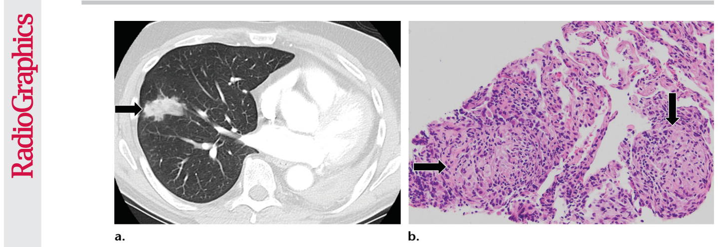
Figure 16. Figure 16. Sarcoid-like granulomatosis of the lung in an asymptomatic 75-year-old man with metastatic melanoma treated with pembrolizumab. (a) Axial CT image shows a parenchymal conglomerate opacity in the right lower lobe of the lung (arrow), which had been gradually increasing over time on serial scans obtained during therapy. Note that the patient had a previous left pneumonectomy. (b) Photomicrograph of specimen from a lung core biopsy of the right lower lobe lesion shows a sarcoid-like reaction characterized by interstitial nonnecrotizing granulomas (arrows) with associated lymphocytic infiltrates. (Hematoxylin- eosin stain; original magnification, 3200.). Gram, silver, and acid-fast bacilli stains (not shown) were negative for microorganisms.
Figure 19
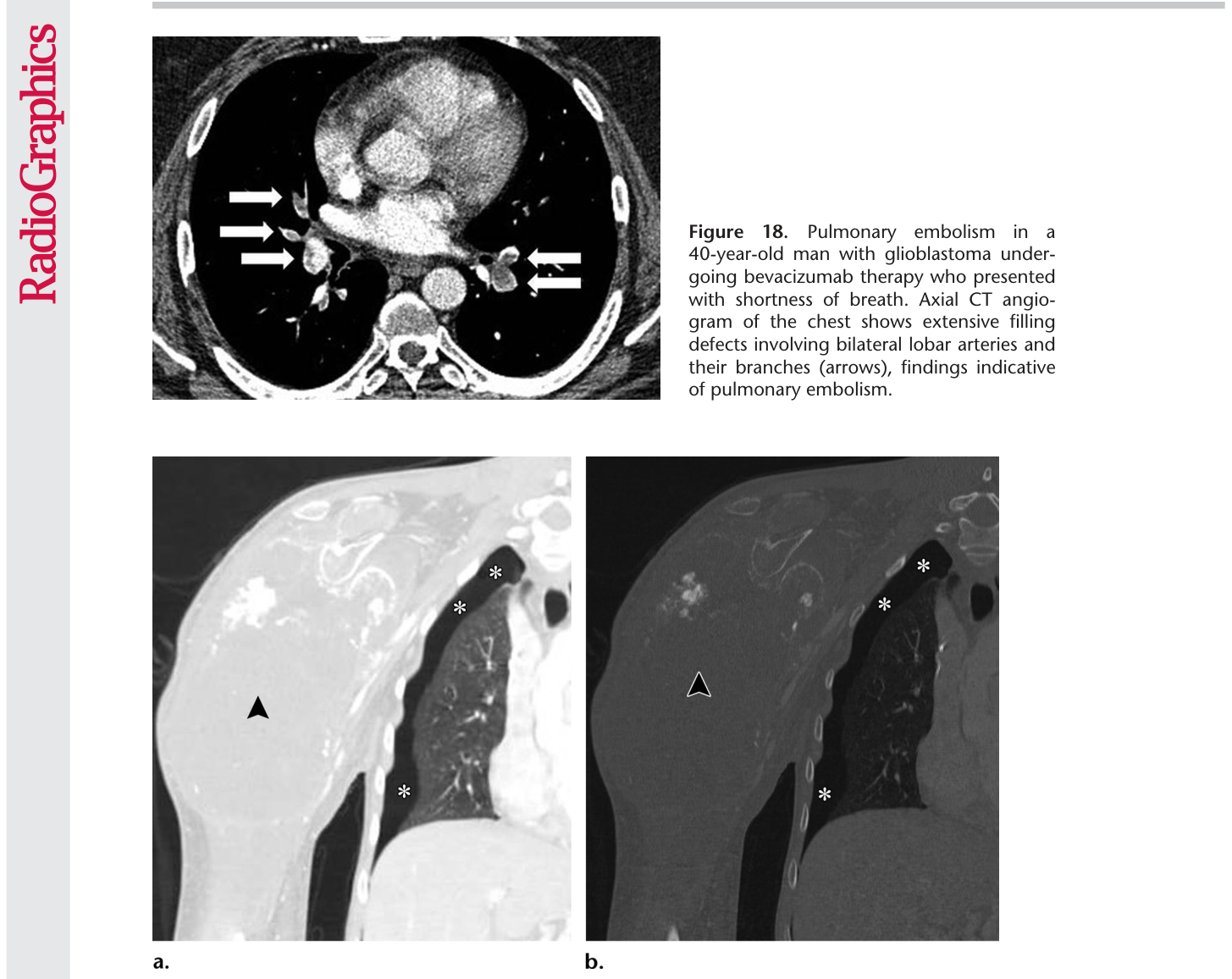
Figure 19. Figure 19. Pneumothorax in a 19-year-old man with metastatic osteosarcoma treated with pazo panib, a multitargeted tyrosine kinase inhibitor. Coronal CT images of the right shoulder with lung window (a) and bone window (b) settings show a large soft-tissue mass (arrowhead) arising from the right humerus. The finding represents primary osteosarcoma metastatic to the lung (not shown). Right pneumothorax (*) is seen and is a known complication of pazopanib therapy.
Figure 20
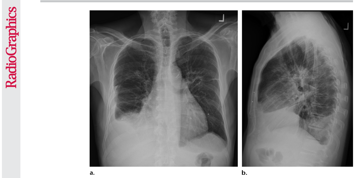
Figure 20. Figure 20. Pleural effusion in a 69-year-old man with a metastatic gastrointestinal stromal tumor treated with nilotinib, a tyrosine kinase inhibitor used for imatinib-resistant chronic myelogenous leukemia. The patient pre sented with increased shortness of breath and bilateral lower extremity edema. Posteroanterior (a) and lateral (b) chest radiographs show bilateral pleural effusion, with bilateral perihilar densities reflective of edema.
Figure 21
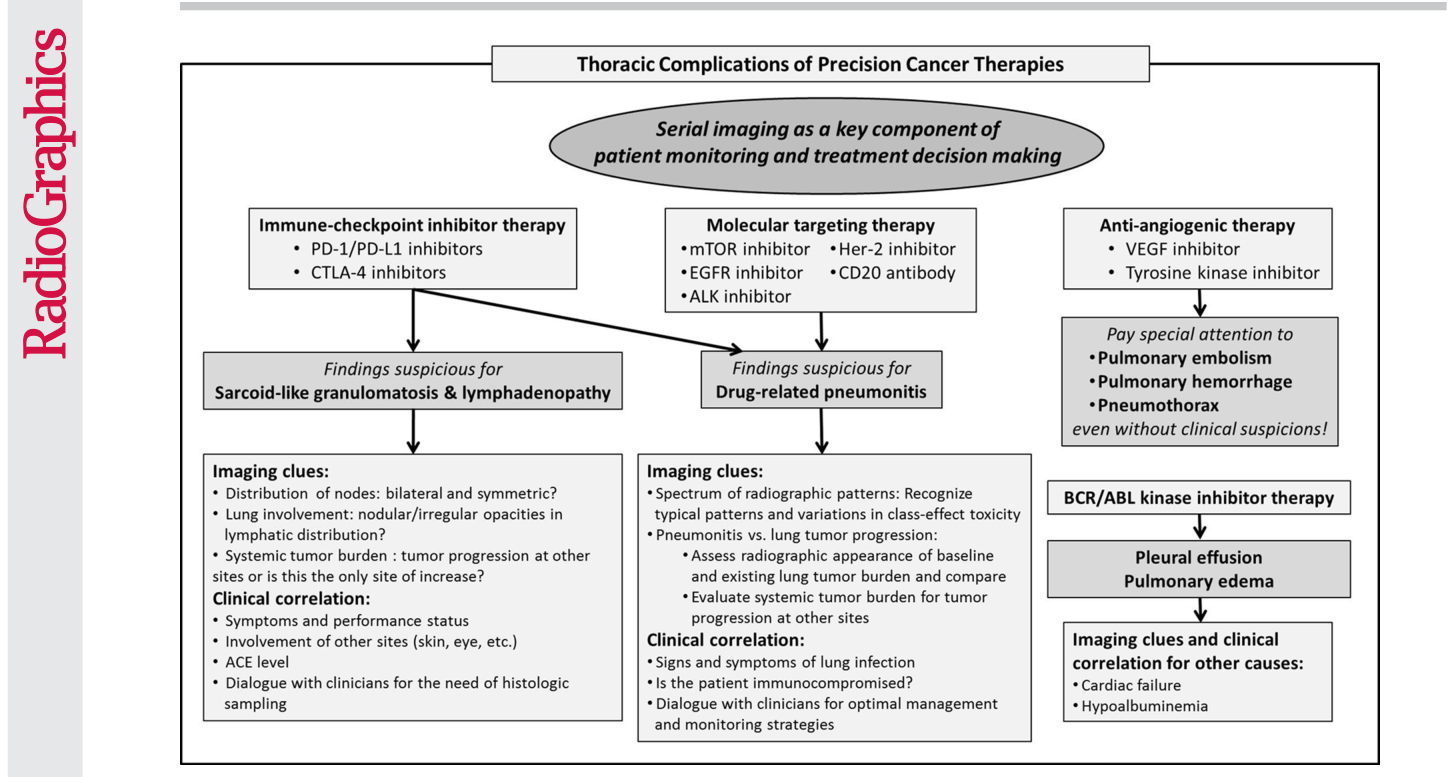
Figure 21. Figure 21. Diagram shows the radiologist’s approach to thoracic complications of precision cancer therapy. A combination of diagnostic clues at imaging and clinical dialogue in the context of precision oncology can contribute to accurate diagnosis and optimal patient care. ACE = angiotensin-converting enzyme.


コメントを残す