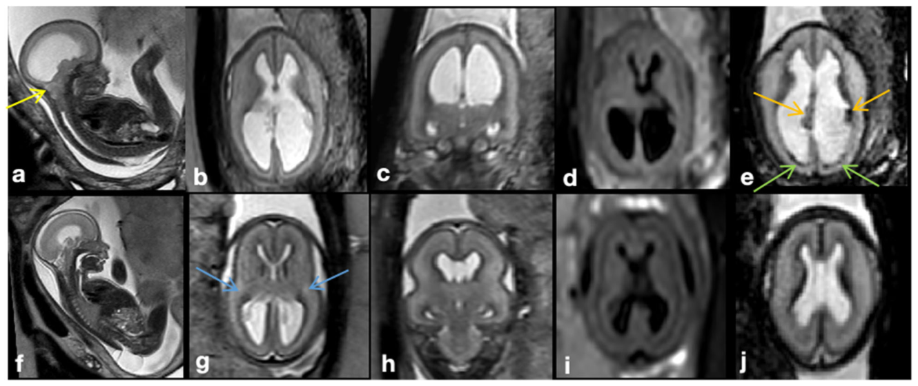
Figure 1. Fig. 1 a–e MRI of a fetus with Chiari II malformation and brain edema at GW24 + 0, spinal defect formation at the lumbar level (L1/2) extending to the sacral level (S1). The posterior cranial fossa was very crowded and the vermis reached up to about C5, accompanied by protrusion of the atlantooccipital membrane (yellow arrow). b T2W-FSE images showed the compressed brain parenchyma with higher signal intensity compared to the age-matched nonedema case (without identifying the parietal crossroads) and effacement of external CSF spaces. c Blurring of lamination was shown on T2-Flair sequences. e Blood-sensitive sequences showed congested, periventricular veins (orange arrow; see Appendix Fig. 2 for postmortem MRI confirmation) and tiny hemorrhages (green arrow) along the ependyma of the posterior horn of the lateral ventricles. f–j MRI of a fetus with Chiari II malformation without brain edema at GW24 + 0. A spinal defect was found at the level of S1. f–h T2W-FSE image showed preserved external CSF spaces and distinguishable hyperintensity of the parietal crossroads in a triangle shape (blue arrows). i Normal brain lamination was shown on T2-Flair sequences. j Blood-sensitive image was normal解説: チアリII奇形と脳浮腫のある胎児(a-e)と脳浮腫のない胎児(f-j)のMRI画像。脳浮腫のある胎児では、後頭蓋窩の混雑、T2強調画像での脳実質の信号強度上昇、外部髄液腔の消失、およびT2-Flairシーケンスでの層構造の不明瞭化が示されている。脳浮腫のない胎児では、外部髄液腔が保たれ、正常な層構造が確認できる。
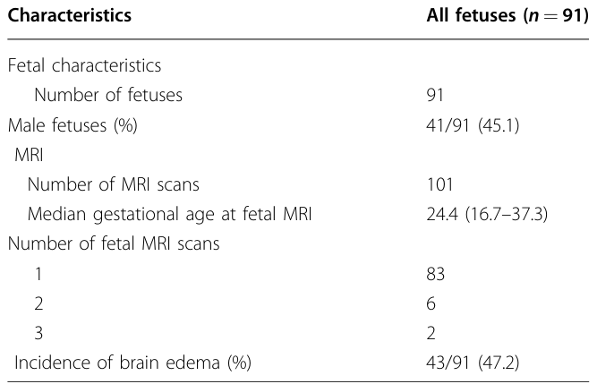
101件のMRIスキャンのうち、50件が脳浮腫ありのチアリII奇形群に分類されました。
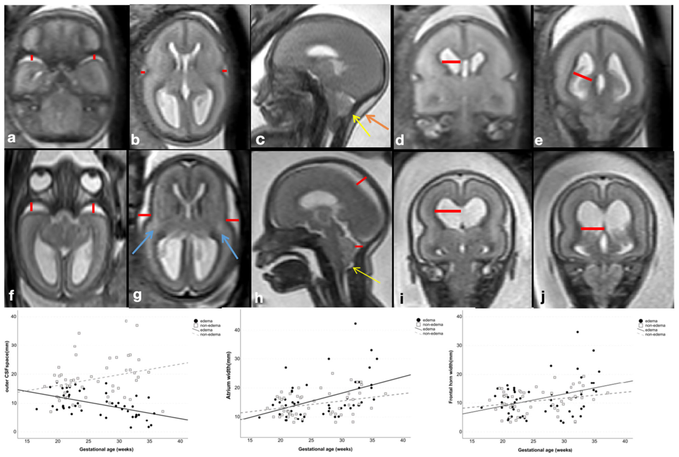
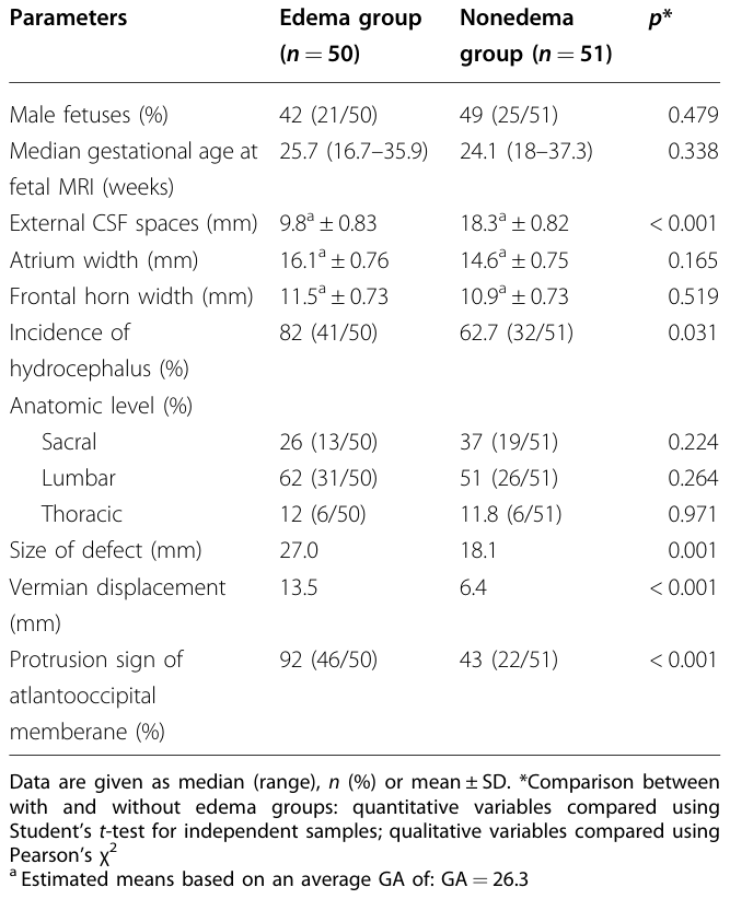
脳浮腫群では、外部髄液腔が有意に減少していることが確認されました(浮腫群9.8mm vs 非浮腫群18.3mm、p < 0.001)。
Figure 2. Fig. 2 Example of external CSF space and frontal horn/atrium width measurement. a–e T2-weighted images of a fetus with Chiari II malformation and edema at GW23 + 3. The brain parenchyma was edematous and swollen, with outer cerebrospinal fluid spaces are only detectable in the insular cistern and temporobasal cistern (axial plane), as well as central and retrocerebellar subarachnoid spaces (mid-sagittal plane), and all the above width measurements (red lines) were summarized as total external CSF space for comparison. The frontal horn and atrium width of the lateral ventricle were also measured. Note the protrusion of the atlantooccipital membrane (yellow arrow) and neck edema (orange arrow). f–j T2-weighted images of an age- matched nonedema fetus at GW23 + 0, external cerebrospinal fluid spaces were preserved, and a triangle-shaped hyperintensity of the parietal crossroads can be 1identified (blue arrows). Protrusion of the atlantooccipital membrane was also presented in this case (yellow arrow). Scatterplot of outer CSF spaces and atrium/frontal horn width throughout the investigated gestational weeks in edema and nonedema groups. A linear decline in outer CSF space width was shown among fetuses investigated in the edema group, with an R2 value of 0.261. GA gestational age given in weeks解説: チアリII奇形と浮腫のある胎児(a-e)と、年齢をマッチさせた非浮腫胎児(f-j)のT2強調画像。外部髄液腔(赤い線)の幅が測定されており、浮腫群では外部髄液腔が著しく減少していることが示されている。下段の散布図は、妊娠週数ごとの外部髄液腔、側脳室前角幅、心房幅の変化を示しており、浮腫群では外部髄液腔幅が妊娠週数の増加とともに直線的に減少する傾向が見られる。
Table 2. Table 2 Fetal and MRI structure characteristics comparisons between with and without edema groups解説: 浮腫群(50件)と非浮腫群(51件)の間で胎児およびMRI構造特性を比較した表。外部髄液腔のサイズ、水頭症の発生率、脊髄欠損のサイズ、小脳虫部の変位、環椎後頭膜の突出などの項目で両群間に有意な差が認められた。
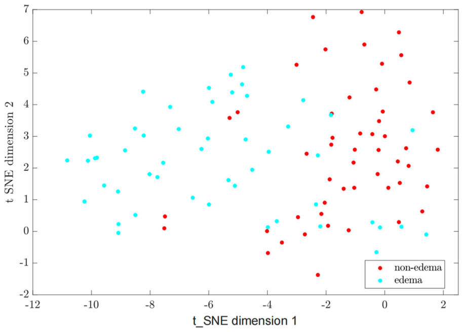
Figure 6. Fig. 6 Two-dimensional t-SNE analysis revealed distinct clustering of the edema and nonedema groups based on the Chiari II malformation-associated features解説: チアリII奇形関連特徴に基づく、浮腫群と非浮腫群の明確なクラスター化を示す2次元t-SNE分析図。浮腫のあるケース(水色)と浮腫のないケース(赤色)が異なる領域に分布していることが視覚的に確認できる。
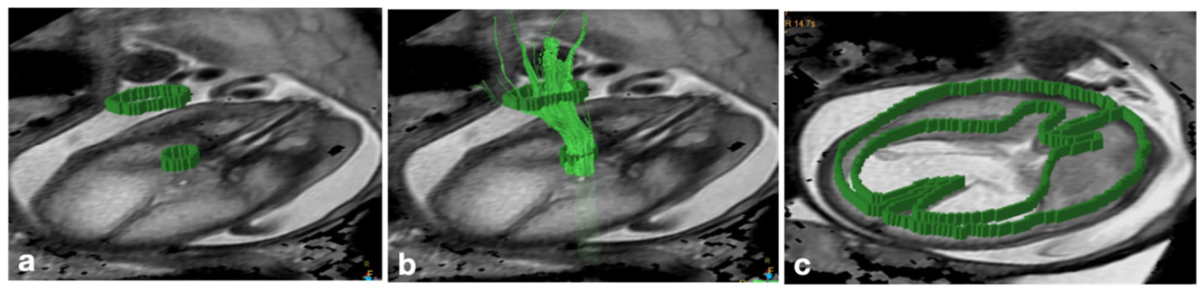
Figure 3. Fig. 3 a, b Characterization of the right corticospinal trajectories by two ROIs, one located at the cerebral peduncles and another at the posterior limb of the internal capsule in a fetus with CM II and brain edema at 28 GW. c A single polygonal ROI was placed at the level of the falx delineating the whole brain parenchyma, to calculate brain parenchyma ADC/FA value解説: チアリII奇形と脳浮腫のある胎児における皮質脊髄路(CST)の軌跡と、脳実質全体の関心領域(ROI)の特性評価を示す。a、bは脳脚と内包後脚に配置された2つのROIによる右皮質脊髄路の軌跡、cは脳鎌のレベルで脳実質全体を囲む単一の多角形ROIを示し、脳実質の見かけの拡散係数(ADC)と分数異方性(FA)値の計算に用いられる。
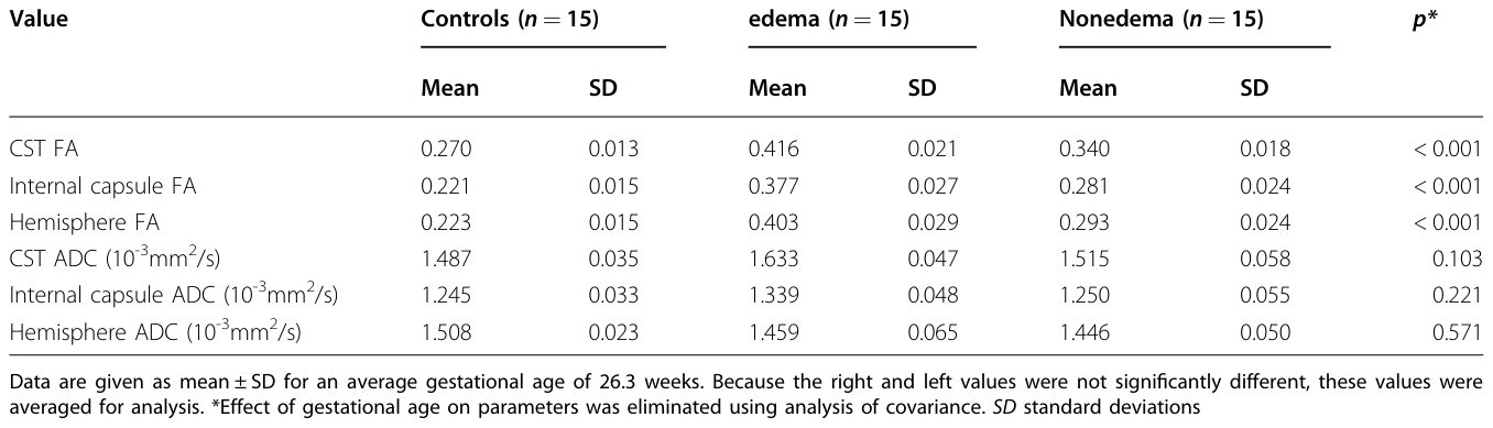
Table 3. Table 3 FA/ADC values in 3 ROIs comparisons between Chiari II malformation with and without edema and normal control groups解説: コントロール群、浮腫群、非浮腫群の3つの関心領域(皮質脊髄路、内包、半球)におけるFA(分数異方性)値とADC(見かけの拡散係数)値を比較した表。浮腫群では、すべての関心領域でFA値が有意に高かったが、ADC値には有意な差は見られなかった。
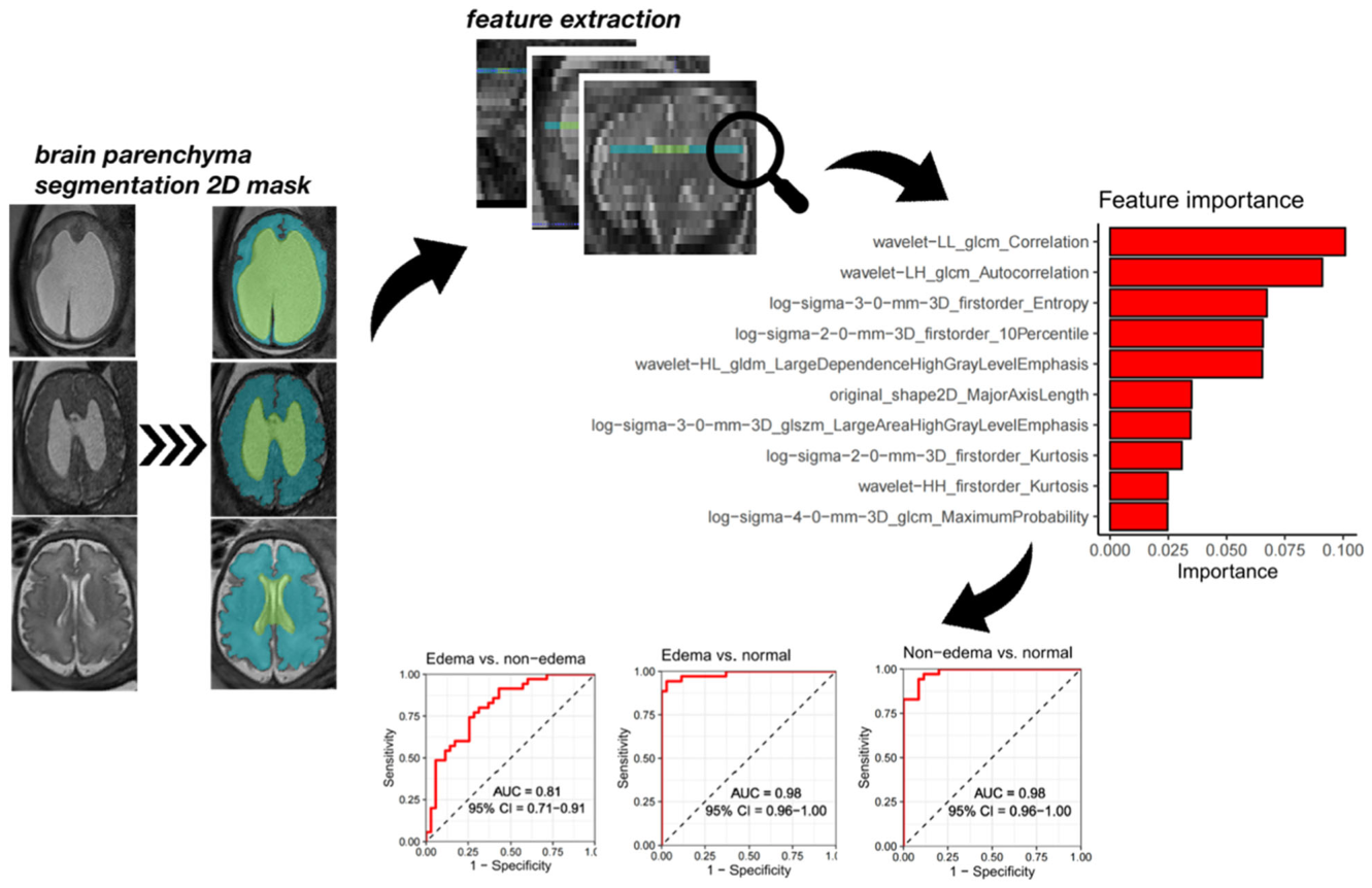
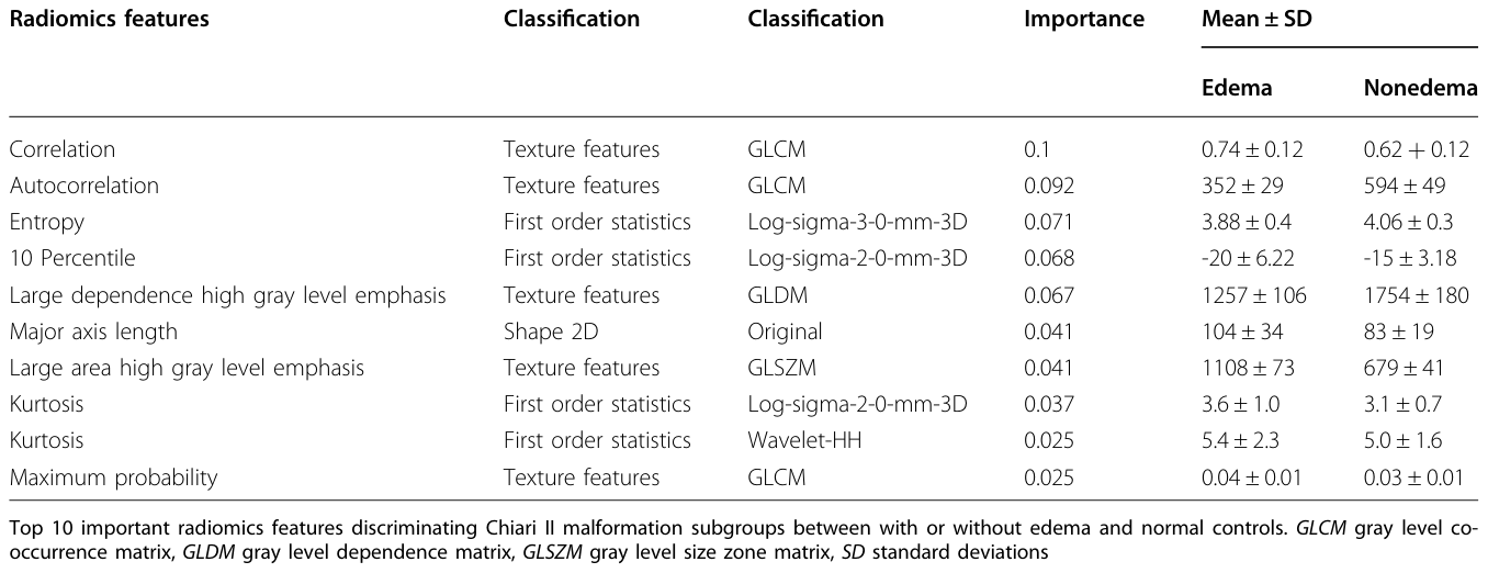
Figure 4. Fig. 4 The diagram shows the workflow for extracting radiomics features from T2W-FSE images. Examples of brain parenchyma segmentation two- dimensional (2D) masks were shown in 3 1:1 age-matched representatives in the edema/nonedema/normal group at GW32 + 3,32 + 3,33, respectively. 2D ROIs were defined at the level of the falx, which was the same level as used in DTI hemisphere segmentation. The ten most important features, which were predominantly texture features, were plotted. The ROC curves of the features showed an AUC of 0.81 (95%CI: 0.71,0.91) for discriminating Chiari II fetuses with edema from those without edema and AUCs of 0.98 (95% CI: 0.96, 1 for both) for discriminating Chiari II cases with and without edema from normal controls. GLCM gray level co-occurrence matrix, GLDM gray level dependence matrix, GLSZM gray level size zone matrix解説: T2W-FSE画像からラジオミクス特徴量を抽出するワークフロー図。脳鎌のレベルで脳実質の2次元セグメンテーションマスクが作成され、そこからラジオミクス特徴が抽出される。重要度の高い上位10の特徴がプロットされ、ROC曲線は浮腫群と非浮腫群の識別においてAUC 0.81、浮腫あり/なし群と正常群の識別においてAUC 0.98を示す。
Table 4. Table 4 The top 10 important radiomics features解説: チアリII奇形サブグループ(浮腫あり/なし)の識別において重要度の高い上位10個のラジオミクス特徴量とその分類、重要度、平均値、標準偏差を示す表。これらの特徴のほとんどはテクスチャ特徴であり、脳の微細構造の変化を反映していると考えられる。
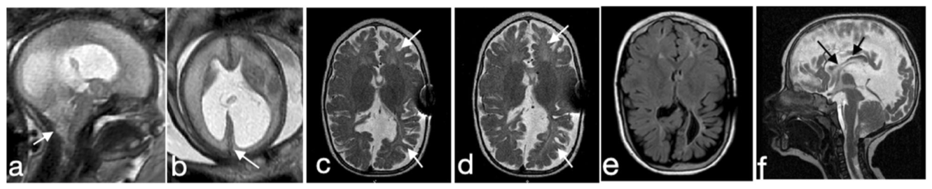
Figure 5. Fig. 5 Postnatal follow-up MRI of a Chiari II malformation with brain edema case. a Prenatal MRI performed at 27 + 3 weeks, showed small posterior fossa, severe vermis ectopia, kinking of the brainstem, and protrusion of the atlantooccipital membrane (white arrow). b Axial plane showed diffuse brain parenchyma edema and compressed superior sagittal sinus (white arrows). c–e Global white matter volume loss and gliosis were shown on postnatal follow-up MRI after shunting conducted at 9 months (c) and 3 years of age (d, e), respectively. f The sagittal plane showed misshaped corpus callosum as a consequence of prenatal compression on the one hand and postnatal shunt implantation on the other hand (black arrows)解説: チアリII奇形と脳浮腫のある胎児の出生前(a, b)および出生後(c-f)のMRI追跡調査画像。出生前の画像では、後頭蓋窩の狭小化、小脳虫部の異所性、脳幹の屈曲、環椎後頭膜の突出が示されている。出生後の画像では、シャント手術後も広範な白質容量減少と膠症(神経組織の瘢痕化)が見られる。

コメントを残す