どうも、Beyond the Pixelです。医療画像診断において、胸壁病変は比較的まれであり、画像上で遭遇すると診断が難しい場合があります。専門家は、他の適応のために行われた検査で偶発的にこれらの病変を発見したり、疑わしい病変を具体的に評価するよう依頼されたりすることがあります。多くの胸壁病変は、画像のみで正確な診断を可能にする特徴的な画像所見を持っていますが、中には所見が大きく重複するため、画像診断のみでの鑑別が困難なものもあります。本記事では、良性病変(「触れてはいけない」とされる、画像のみで診断可能で、症状がない限り生検や切除が不要なもの)と、確実に特徴づけられず、さらなる精査が必要な病変の両方の画像特徴について解説します。
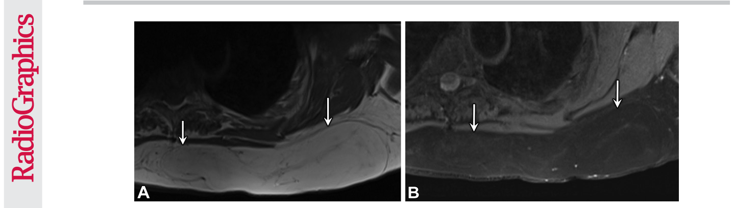
Figure 1. Figure 1. Lipoma in a 59-year-old woman who presented with a left chest mass that had been growing during the past year. Contrast-enhanced MRI of the chest was performed. Transaxial non–fat-saturated T1-weighted MR image (A) shows a hyperintense mass (arrows) that is nulled on the transaxial fat-saturated T1-weighted MR image (B). The lesion showed no enhancement on contrast-enhanced MR images (not shown). Owing to its growth, the mass was resected, and pathologic analysis confirmed lipoma.解説: 59歳女性の左胸部腫瘤における脂肪腫の画像です。非脂肪抑制T1強調MR画像(A)では高信号の腫瘤が確認され、脂肪抑制T1強調MR画像(B)では信号が抑制されています。これは脂肪組織で構成される良性腫瘍である脂肪腫の特徴を示しています。
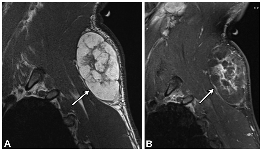
Figure 2. Figure 2. Spindle cell lipoma in a 42-year-old man who presented with a lump in the up- per left chest wall. (A) Sagittal non–fat-saturated T1-weighted MR image shows a fat-con- taining mass (arrow) with multiple thick hypointense septa, which were also hyperintense at T2-weighted MRI (not shown). (B) Sagittal contrast-enhanced T1-weighted fat-saturated MR image shows enhancement of the thick septa in the mass (arrow). The mass was excised owing to concern for liposarcoma, and spindle cell lipoma was found.解説: 42歳男性の左上胸壁のしこりにおける紡錘細胞脂肪腫の画像です。非脂肪抑制T1強調MR画像(A)では脂肪含有性の腫瘤に複数の厚い低信号隔壁が見られ、造影T1強調脂肪抑制MR画像(B)ではこれらの厚い隔壁に造影増強が認められます。
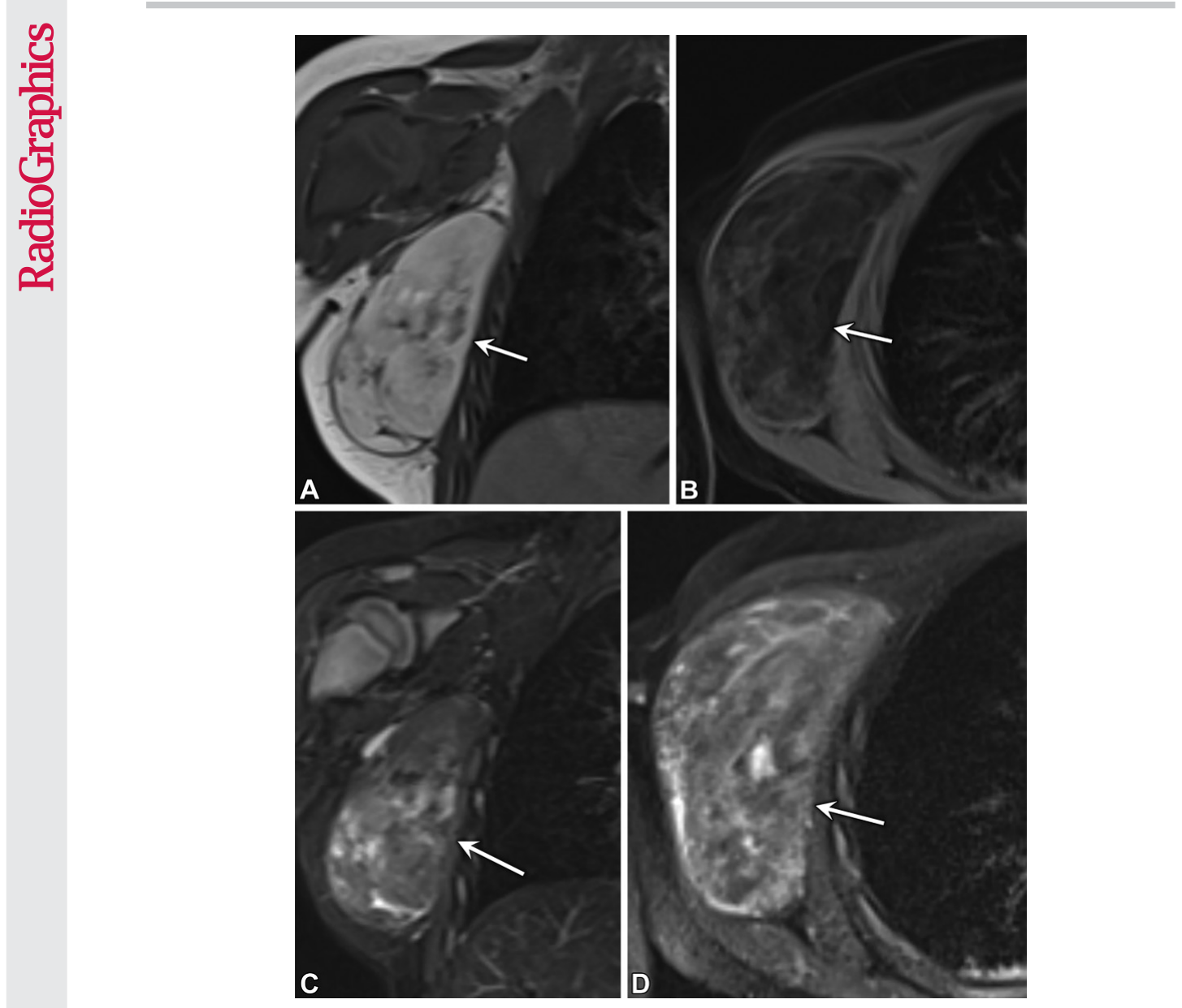
Figure 3. Figure 3. Lipoblastoma in a 2-year-old boy who had a palpable mass in the right axilla and underwent contrast-enhanced MRI. (A, B) Coronal T1-weighted non–fat-saturated im- age (A) shows areas of hyperintensity (arrow), which are suppressed on the transaxial T1- weighted fat-saturated image (B). (C, D) Coronal T2-weighted short τ inversion-recovery image (C) shows multiple hyperintense septa in the mass (arrow), with corresponding en- hancement on the transaxial contrast-enhanced T1-weighted fat-saturated image (D). The mass was resected, and pathologic analysis revealed lipoblastoma.解説: 2歳男児の右腋窩の触知可能な腫瘤における脂肪芽腫の画像です。冠状T1強調非脂肪抑制画像(A)では高信号領域が見られ、軸状T1強調脂肪抑制画像(B)ではこれが抑制されています。冠状T2強調STIR画像(C)では腫瘤内に複数の高信号隔壁が示され、軸状造影T1強調脂肪抑制画像(D)では対応する増強が認められます。
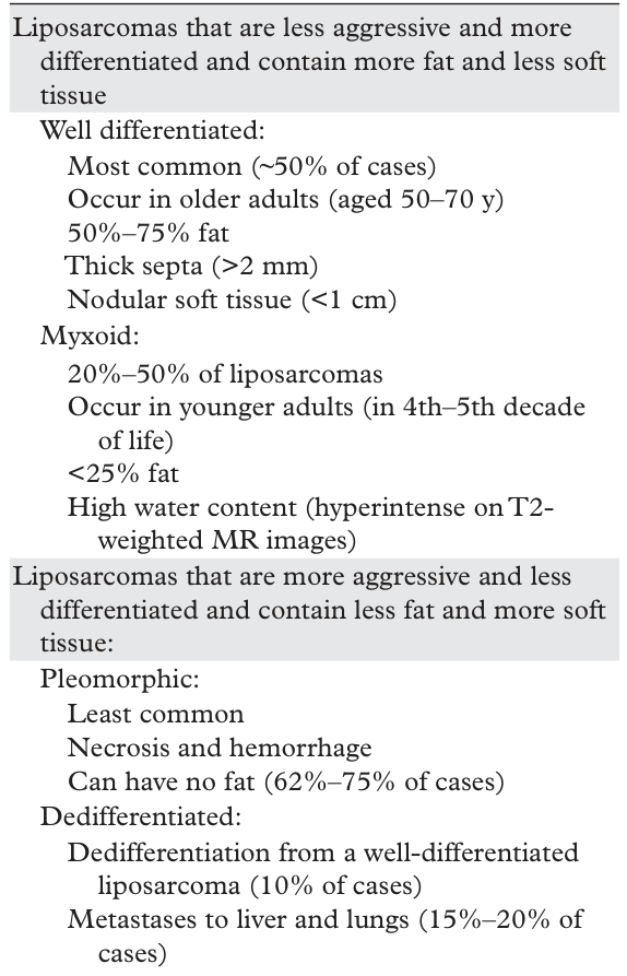
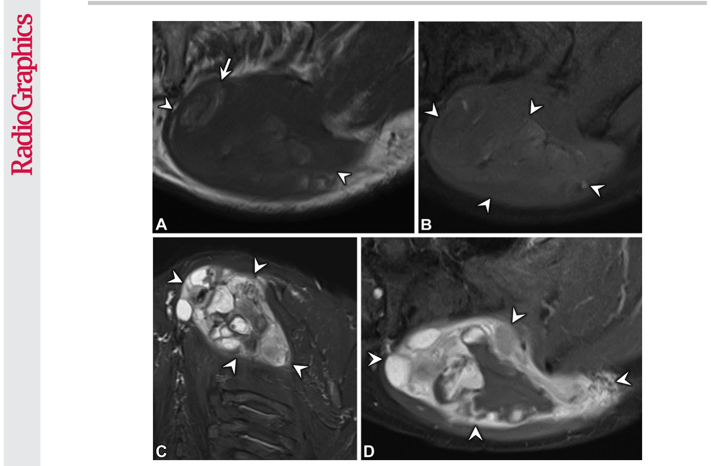
Figure 4. Figure 4. Myxoid liposarcoma in a 72-year-old man with a palpable mass involving the left upper back. (A, B) Transaxial non–fat-saturated T1-weighted MR image (A) shows a T1-hypointense mass (ar- rowheads) with areas of T1 hyperintensity (arrow in A), which are null on the transaxial fat-saturated T1-weighted MR image (B). (C) Coronal short τ inversion-recovery MR image shows areas of very hy- perintense T2 signal, representing the myxoid areas of the lesion (arrowheads). (D) Transaxial contrast- enhanced fat-saturated T1-weighted MR image shows avid enhancement of the T2-hyperintense areas of the mass (arrowheads). The mass was resected, and histologic analysis confirmed myxoid liposarcoma.解説: 72歳男性の左上背部腫瘤における粘液型脂肪肉腫の画像です。軸状非脂肪抑制T1強調MR画像(A)ではT1低信号の腫瘤(矢頭)とT1高信号領域(Aの矢印)が見られ、脂肪抑制T1強調MR画像(B)ではT1高信号領域が抑制されています。冠状STIR MR画像(C)では、病変の粘液領域を表す非常に高信号のT2信号領域が示されています。軸状造影脂肪抑制T1強調MR画像(D)ではT2高信号領域の著明な増強が認められます。
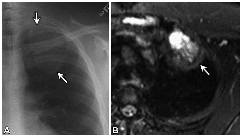
Figure 5. Figure 5. Fibrous dysplasia in a 37-year-old man. (A) Posteroanterior chest radiograph shows an incidentally discovered expansile mass (arrows) involving the left second rib, with ground- glass attenuation. (B) Transaxial T2-weighted fat-saturated MR image shows a hyperintense mass (arrow) in the left second rib that was hypointense at T1-weighted MRI (not shown). The mass was resected after biopsy revealed a spindle neoplasm. Findings at histologic analysis after resection confirmed fibrous dysplasia.解説: 37歳男性の線維性異形成の画像です。後前部胸部X線写真(A)では、偶発的に発見された左第2肋骨の膨張性腫瘤が示され、すりガラス状の吸収を呈しています。軸状T2強調脂肪抑制MR画像(B)では、左第2肋骨に高信号の腫瘤が確認され、T1強調MR画像では低信号でした。
Figure 6. Figure 6. Osteochondroma found at lung cancer screening in a 66-year-old woman who smokes. Axial noncontrast CT image shows an osteochondroma (ar- row) from the anterior left fourth rib with corticomedullary continuity.解説: 66歳女性の肺癌スクリーニングで発見された骨軟骨腫の画像です。軸状非造影CT画像では、左第4肋骨前方から皮質骨髄連続性を伴って発生する骨軟骨腫が示されています。
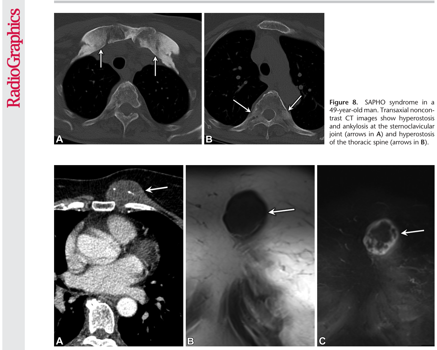
Figure 9. Figure 9. Chondrosarcoma in a 71-year-old woman. (A) Contrast-enhanced chest CT image obtained to assess a painful chest lump shows a mass (arrow) arising from the left anterior costochondral junction. The mass has central, punctate, and archlike calcifications (chondroid matrix). (B) Coronal noncontrast non–fat-saturated T1-weighted MR image shows a hypointense mass (arrow). (C) Coro- nal contrast-enhanced fat-saturated MR image shows peripheral nodular enhancement (arrow). Histopathologic analysis confirmed the diagnosis of chondrosarcoma.解説: 71歳女性の軟骨肉腫の画像です。疼痛を伴う胸部腫瘤を評価するために取得された造影胸部CT画像(A)では、左前肋軟骨接合部から発生する腫瘤が示され、中心に点状および弓状の石灰化(軟骨様基質)があります。冠状非造影非脂肪抑制T1強調MR画像(B)では低信号の腫瘤が確認され、冠状造影脂肪抑制MR画像(C)では周辺結節性増強が認められます。
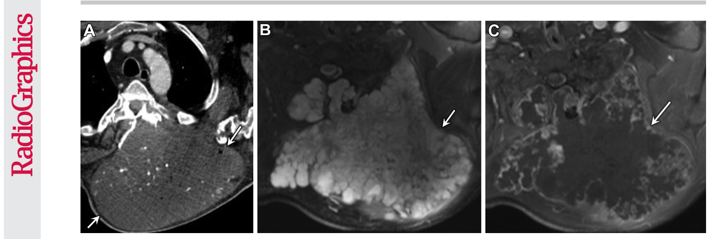
Figure 11. Figure 11. Chondrosarcoma arising from osteochondroma in a 57-year-old man with a history of hereditary osteochondromas who presented with a left upper back mass that had been slowly growing for the past 10 years. (A) Transaxial contrast-enhanced chest CT image shows a large mass (arrows) with a stippled chondroid matrix arising from an osteochondroma. The small locules of gas are secondary to biopsies performed the previous day. (B) Transaxial T2-weighted fat-saturated MR image shows a hyperintense mass (arrow). (C) Transaxial contrast-enhanced T1-weighted fat-saturated MR image shows peripheral nodular enhancement (arrow). Biopsy findings confirmed a low-grade chondrosarcoma.解説: 遺伝性骨軟骨腫の病歴を持つ57歳男性の骨軟骨腫から発生した軟骨肉腫の画像です。軸状造影胸部CT画像(A)では、骨軟骨腫から発生する、点状の軟骨様基質を伴う大きな腫瘤が示されています。軸状T2強調脂肪抑制MR画像(B)では高信号の腫瘤が確認され、軸状造影T1強調脂肪抑制MR画像(C)では周辺結節性増強が認められます。
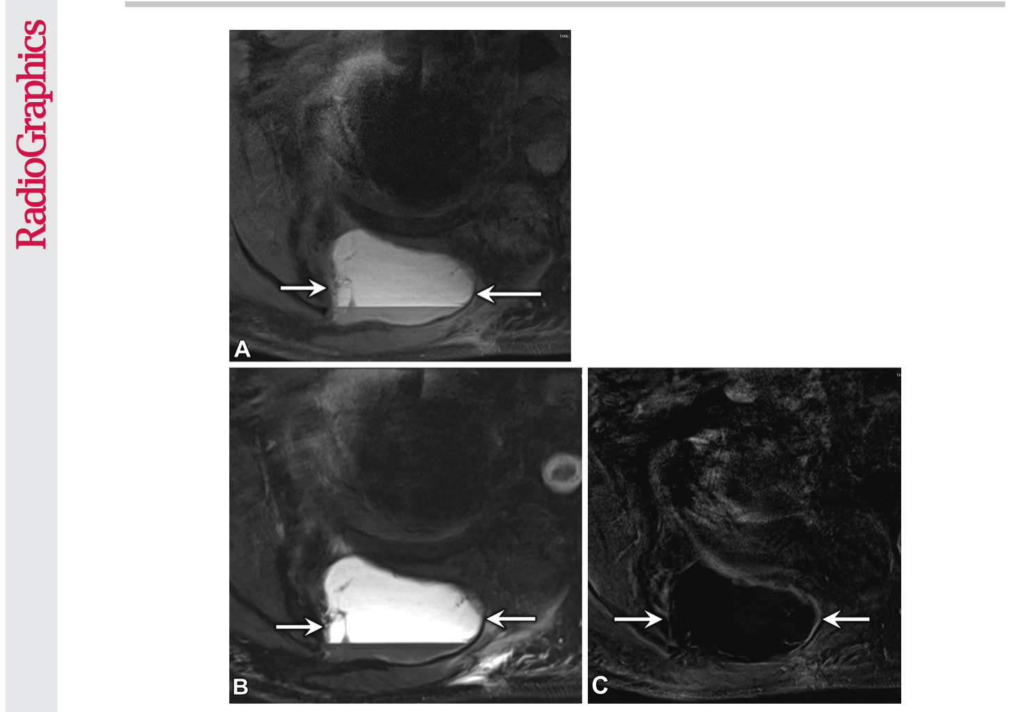
Figure 22. Figure 22. Hematoma in a 58-year-old man who underwent transaxial MRI after resection of a poste- rior chest wall desmoid tumor. (A) Noncontrast T1-weighted fat-saturated image shows a hyperintense fluid collection (arrows) with a fluid-fluid level at the resection site. (B) T2-weighted fat-saturated image shows the fluid collection (arrows), and similar to A, a relatively hypointense dependent layer. (C) Post- contrast fat-saturated T1-weighted subtraction image shows no enhancement (arrows), consistent with a hematoma. The collection was resolved at 6-month follow-up MRI.
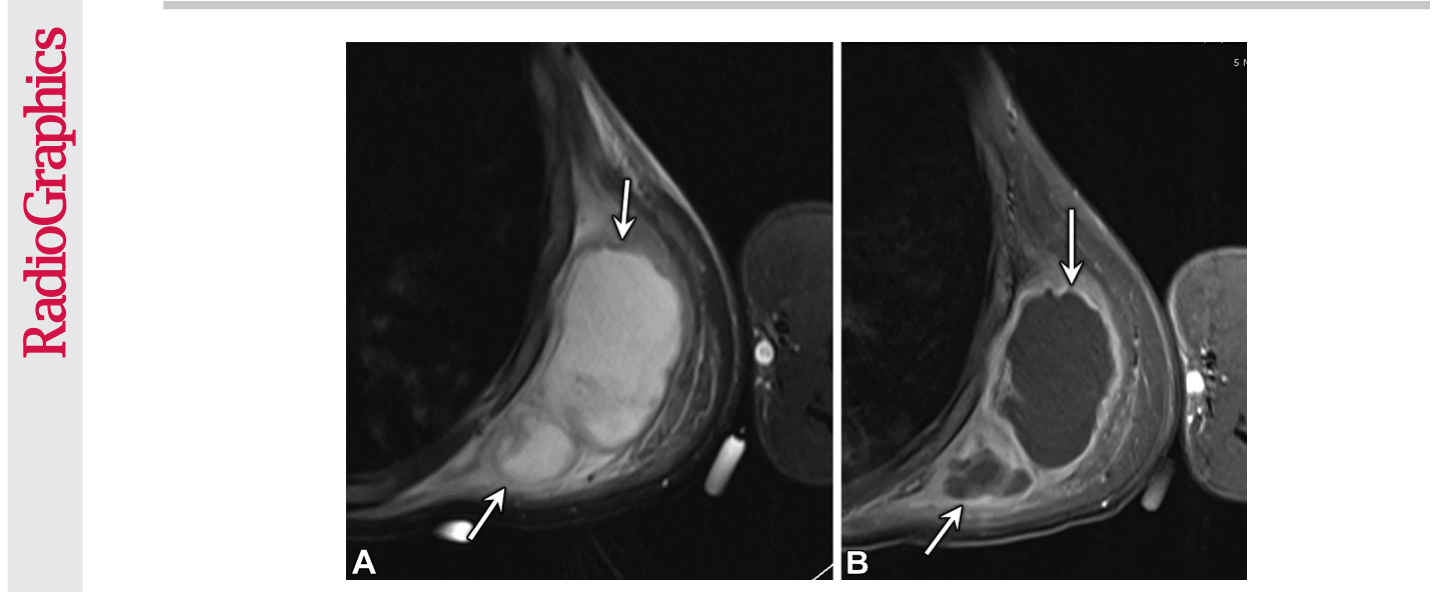
Figure 23. Figure 23. Abscess in a 33-year-old man with a history of common variable immunode- ficiency who presented with fever and swelling. (A) Transaxial T2-weighted MR image re- veals a loculated fluid collection (arrows) in the left chest wall. (B) Transaxial postcontrast T1-weighted fat-saturated MR image shows a thick rind of peripheral enhancement (arrows). Abscess was confirmed at surgical debridement.
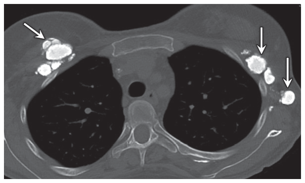
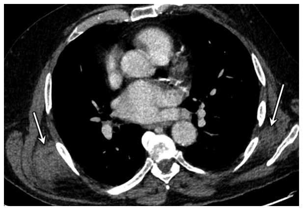
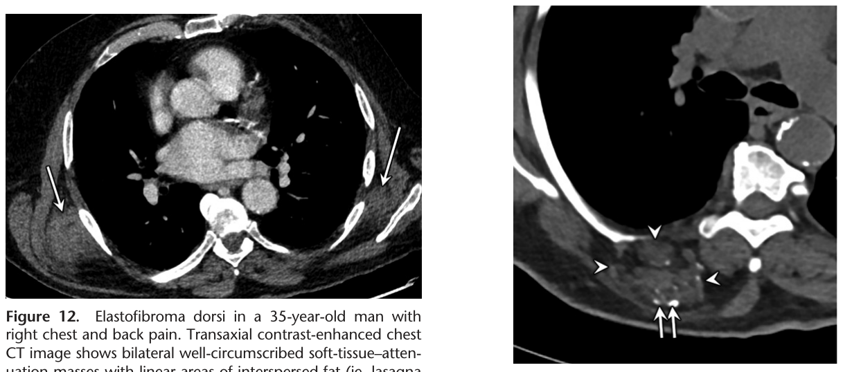
Figure 12. Figure 12. Elastofibroma dorsi in a 35-year-old man with right chest and back pain. Transaxial contrast-enhanced chest CT image shows bilateral well-circumscribed soft-tissue–atten- uation masses with linear areas of interspersed fat (ie, lasagna sign) (arrows) in the posterior chest wall, deep to the latissi- mus dorsi and anterior to the scapular tip. The right mass was causing pain and discomfort and thus was removed. Histologic analysis confirmed elastofibroma dorsi.
Figure 13
Figure 13. Figure 13. Hemangioma in a 73-year-old woman who underwent noncontrast CT before coronary artery bypass graft placement. Trans- axial CT image shows a soft-tissue lesion (arrow- heads) in the right posterior chest wall, with in- terspersed areas of internal fat and calcifications (arrows), representing phleboliths. when the lesions are bilateral, with attenuation and texture similar to those of skeletal muscle. Elastofibroma dorsi lesions may be mildly T2 hyperintense, particularly on fat-suppressed MR images. Elastofibroma dorsi typically has linear areas of fat within the tumor, which are some- times termed the lasagna sign because of the layering appearance (Fig 12) (41). It should be noted that while the area between the serratus anterior and the scapula is the most common location of elastofibroma dorsi lesions, they may occur less commonly at other sites in the tho- rax, with the second most common site being posterior to the first or second rib and deep to the superior scapula (42). The lesion may show very mild FDG uptake and mild enhancement (41). Elastofibroma dorsi is considered a “do
Figure 14
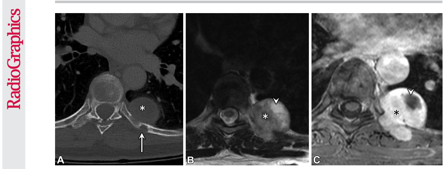
Figure 14. Figure 14. Schwannoma in a 59-year-old woman found to have a left posterior chest wall mass at CT performed for gallstone pan- creatitis. (A) Transaxial contrast-enhanced CT image (bone window) shows the mass (*) adjacent to the left eighth rib, with a small area of bone remodeling (arrow). (B) Transaxial T2-weighted MR image shows a hyperintense bilobar mass (*) with a cystic area (arrowhead). (C) Transaxial contrast-enhanced T1-weighted fat-saturated MR image shows avid enhancement of the mass (*) , with no enhancement of the cystic area (arrowhead). Postresection histologic analysis confirmed a schwannoma.
Figure 15
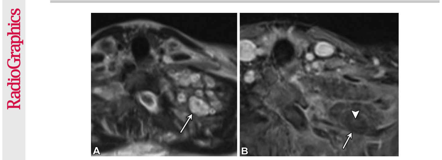
Figure 15. Figure 15. Known neurofibromatosis type 1 in a 19-year-old woman who underwent follow-up MRI for plexiform neurofibromas. (A) Transaxial T2-weighted MR image shows the target sign, with central low signal intensity and high peripheral signal intensity (arrow). (B) Transaxial contrast-enhanced T1- weighted fat-saturated MR image shows a plexiform neurofibroma (arrow), with enhancement of the central portion (arrowhead).
Figure 17
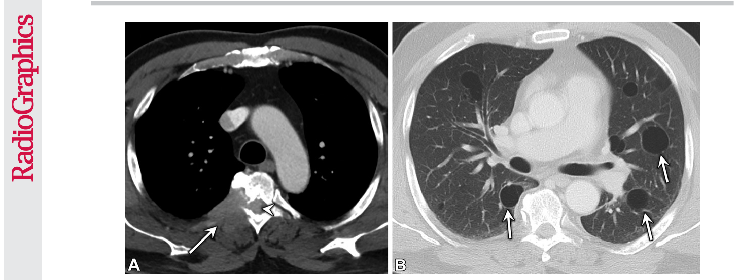
Figure 17. Figure 17. Myeloma in a 69-year-old man with lower extremity weakness. (A) Transaxial contrast-enhanced chest CT image (soft-tissue window) shows a right posterior chest wall mass (arrow) extending into the central canal (ar- rowhead). (B) Transaxial contrast-enhanced CT image (lung window) shows cysts (arrows) in both lungs secondary to amyloid involvement. Results of biopsy of the paraspinal mass confirmed multiple myeloma.
Figure 18
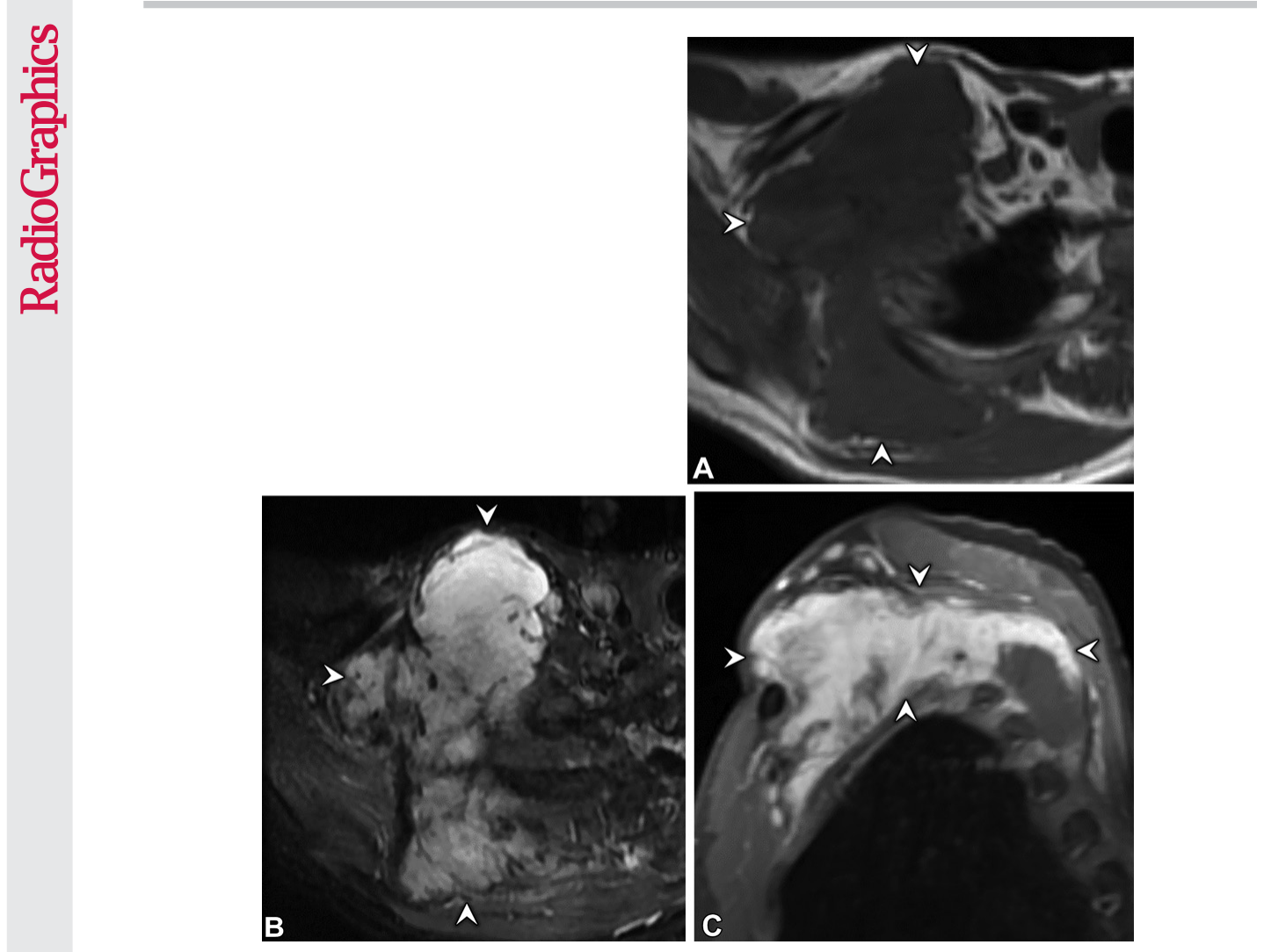
Figure 18. Figure 18. Aggressive fibromatosis (desmoid tumor) in a 55-year-old man with a history of Li-Fraumeni syndrome who presented with right shoulder pain. (A, B) Transaxial noncontrast T1-weighted (A) and short τ inversion-recovery (B) MR images show a T1-isointense (arrowheads in A) and T2-hyperintense (arrowheads in B) mass centered in the right shoulder, with invasion of the right scapula and right second and third ribs. (C) Coronal contrast-enhanced T1-weighted fat-saturated MR image shows avid enhancement (arrowheads). After multiple nondiagnostic biopsies, the mass was partially resected, and histopathologic analysis revealed a desmoid tumor.
Figure 19
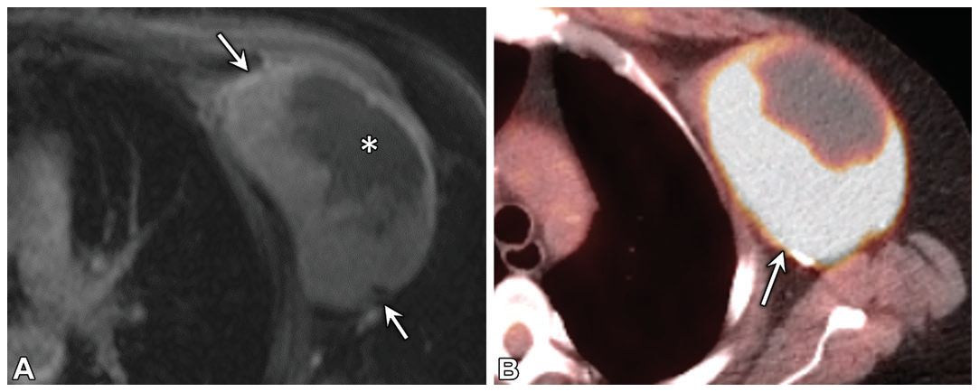
Figure 19. Figure 19. Undifferentiated pleomorphic sarcoma in a 59-year-old woman with a history of left breast cancer 22 years earlier and chemoradiation who presented with a palpable chest wall mass. (A) Transaxial contrast- enhanced T1-weighted fat-saturated MR image shows a mass (arrows) in the left subpectoral chest wall, with a cystic area (*), likely representing necrosis. (B) Transaxial PET/CT image obtained later shows avid FDG uptake (arrow). Biopsy findings confirmed high-grade undifferentiated pleomorphic sarcoma.



コメントを残す