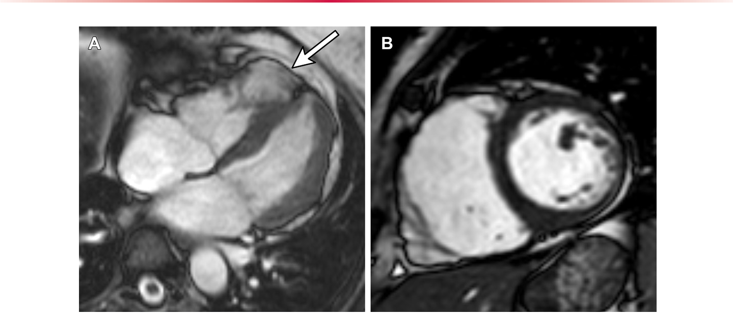
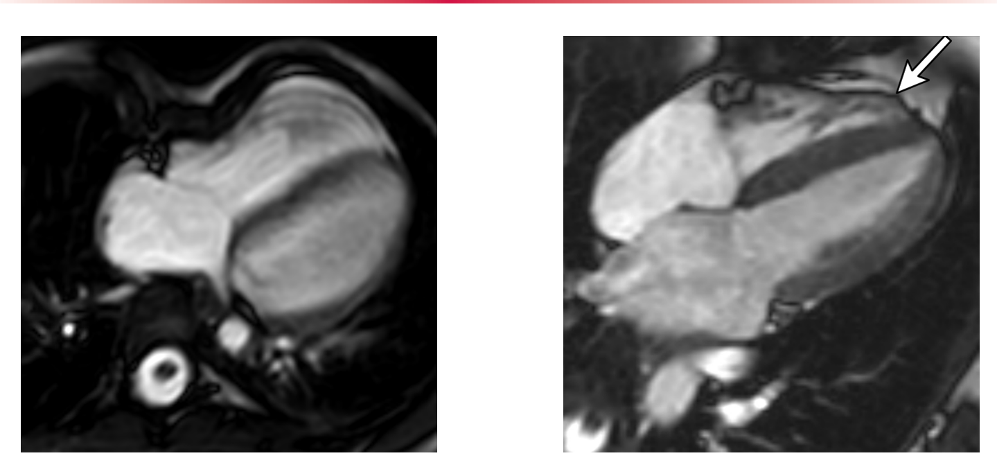
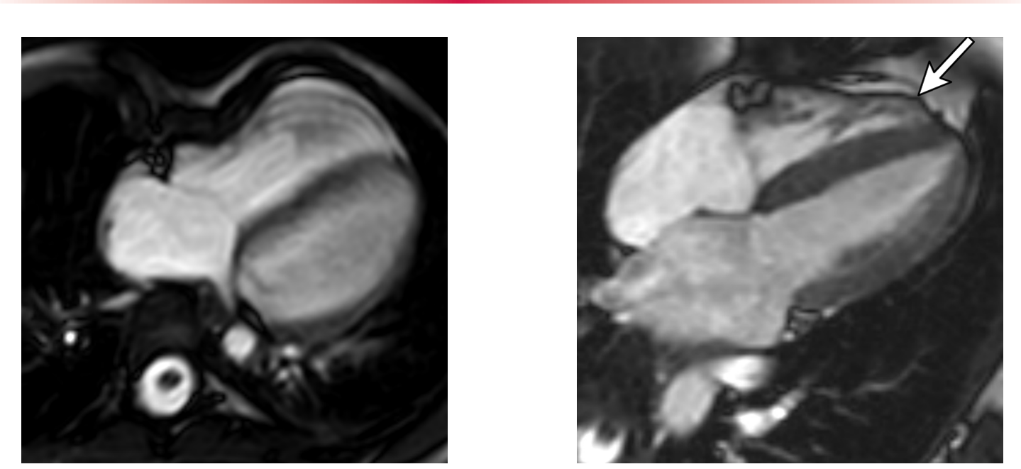
Figure 3. Figure 3. ARVC in a 58-year-old woman who presented with an episode of syncope while exercising on a treadmill. (A) Still frame from cine steady-state free precession (SSFP) cardiac MRI in the horizontal longitudi- nal axis plane shows a focal bulge of the apical segment of the RV free wall (arrow) during systole, consistent with dyskinesis. (B) Still frame from cine SSFP cardiac MRI in a short-axis plane shows RV dilatation (end-dia- stolic indexed volume, 128 mL/m2) with decreased systolic function (ejection fraction, 32%), therefore fulfill- ing one major 2010 task force imaging criterion. The patient was found to have a PKP2 gene variant, therefore satisfying two major criteria, consistent with a definite diagnosis of ARVC.解説: 58歳の女性のARVCの心臓MRI画像で、運動中に失神を経験しました。(A)シネSSFP心臓MRIの水平長軸像(静止画)では、右心室自由壁の心尖部セグメントに局所的な膨隆(矢印)が示され、異常運動と一致します。(B)シネSSFP心臓MRIの短軸像(静止画)では、右心室の拡張(体表面積で補正した拡張末期容量128mL/m2)と収縮機能の低下(駆出率32%)が示されており、2010年タスクフォース画像診断基準の主要基準を1つ満たしています。この対象者にはPKP2遺伝子変異が見つかり、2つの主要基準を満たしたことで、ARVCの確定診断となりました。
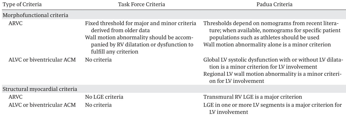
Table 3. Table 3. Key Differences between the 2010 Task Force and Padua Criteria for the Diagnosis of ACM解説: 2010年タスクフォース基準とパドヴァ基準の主な違いを示す表です。形態機能基準において、ARVCのタスクフォース基準は固定された閾値を用いるのに対し、パドヴァ基準はノモグラムに依存します。ALVCまたは両心室型ACMの基準はタスクフォース基準にはないものの、パドヴァ基準には存在します。構造的心筋基準では、タスクフォース基準にLGEの基準はないのに対し、パドヴァ基準では透過性RV LGEが主要基準として含まれています。
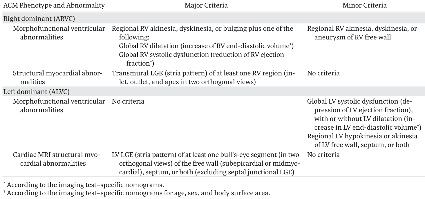
Table 4. Table 4. Padua Criteria for the Diagnosis of ACM with Cardiac MRI解説: パドヴァ基準に基づくACMの心臓MRI診断基準を示す表です。右室優位型(ARVC)では形態機能的異常と構造的心筋異常の主要および副次基準が示されており、ALVCでは形態機能的異常の副次基準と、LV LGEの主要構造基準が示されています。
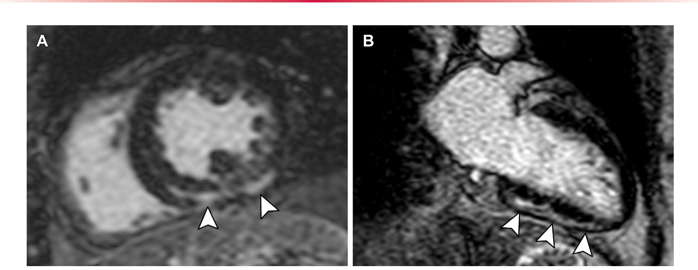
Figure 11. Figure 11. DSP pathogenic variant discovered during family screening in a 53-year-old woman. (A) LGE cardiac MR image in the short-axis plane at the midventricular level shows subepicardial delayed enhancement of the inferior wall and midmyocardi- al delayed enhancement of the inferoseptal wall (arrowheads). (B) LGE cardiac MR image in the vertical longitudinal axis shows that the subepicardial enhancement at the inferior wall extends from the base to the apex of the LV (arrowheads). The cardiac MRI finding is a major criterion, and together with the presence of the genetic mutation, fulfills two major criteria and confirms the diagnosis of ALVC.解説: 53歳の女性において家族スクリーニングで発見されたDSP病原性変異を示す画像です。(A) 心室中隔レベルの短軸LGE心臓MR画像では、下壁の心外膜下遅延増強と、下中隔壁の心筋中央部遅延増強(矢印)が示されています。(B) 垂直長軸LGE心臓MR画像では、下壁の心外膜下増強が左心室の基部から心尖部まで広がっていることが示されています(矢印)。心臓MRI所見は主要基準であり、遺伝子変異の存在と合わせて2つの主要基準を満たし、ALVCの診断を確定しました。
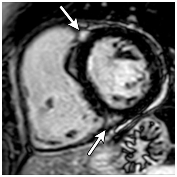
Figure 12. Figure 12. LGE cardiac MR image in the short-axis plane at the midventricular level shows midmyocardial delayed enhancement (arrows) predominantly at the RV septal insertion sites, which can be seen in healthy individuals.解説: 心室中隔レベルの短軸LGE心臓MR画像で、主に右心室中隔挿入部位に心筋中央部遅延増強(矢印)が示されています。これは健康な対象者にも見られる所見です。
Figure 5. Figure 5. Tethering of the RV to the sternum in a 66-year-old man. Still images from cine SSFP cardi- ac MRI in the horizontal long-axis plane at end-diastole (A) and end-systole (B) show tethering (arrow) of the RV free wall to the posterior aspect of the sternum (adjacent to sternal wires). This can mimic regional wall motion abnormalities of the RV.解説: 66歳男性の胸骨への右心室繋留を示すシネSSFP心臓MRI画像です。拡張末期(A)と収縮末期(B)の水平長軸像(静止画)では、右心室自由壁が胸骨後方(胸骨ワイヤーに隣接)に繋留(矢印)されていることが示されています。これは右心室の局所壁運動異常と誤認される可能性があります。⚠ 自動抽出画像の検証で一致を確認できませんでした。正確な内容は元論文のFigure 5をご参照ください。
Figure 6. Figure 6. Pectus excavatum in a 47-year-old man. Axial MR image in a patient shows distortion of the RV, with compression of the RV base and a consequent relative enlargement of the RV apex. This can make assessment of wall motion abnormalities as well as contouring RV volumes challenging.解説: 47歳男性の漏斗胸を示す軸位MRI画像です。右心室の歪みが示され、右心室基部の圧迫とそれに伴う右心室心尖部および右心室流出路の相対的な拡大が見られます。これは壁運動異常の評価や右心室体積の輪郭付けを困難にする可能性があります。
モデレーターバンドと呼ばれる筋肉組織の挿入部が膨隆や壁運動異常と誤解されることもあります。
Figure 7. Figure 7. Apparent abnormality in a 28-year-old woman with a history of type I atrioventricular block. Still image from horizontal longitudinal axis cine SSFP cardiac MRI shows an apicolateral bulge (arrow) adjacent to the insertion of the moderator band in the RV free wall, a finding that can be seen in healthy individuals.解説: 1型房室ブロックの既往がある28歳女性の心臓MRI画像です。水平長軸シネSSFP心臓MRIの静止画は、右心室自由壁のモデレーターバンド挿入部に隣接する心尖部外側の膨隆(矢印)を示しています。これは健康な対象者にも見られる所見です。
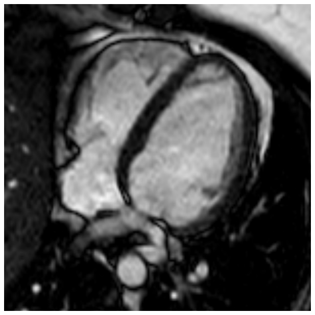
Figure 8. Figure 8. Anatomic variant in a 28-year-old man with a history of premature ventricular contractions. Still image from cine SSFP cardiac MRI in the horizontal longitudinal axis plane shows the LV and RV forming separate cardiac apices, an anatomic variant known as a butterfly apex, which can be seen in healthy individuals.解説: 心室性期外収縮の既往がある28歳男性の解剖学的変異を示す心臓MRI画像です。水平長軸シネSSFP心臓MRIの静止画は、左心室と右心室が別々の心尖部を形成している様子を示しており、「蝶形心尖部」として知られる解剖学的変異です。これは健康な対象者にも見られます。
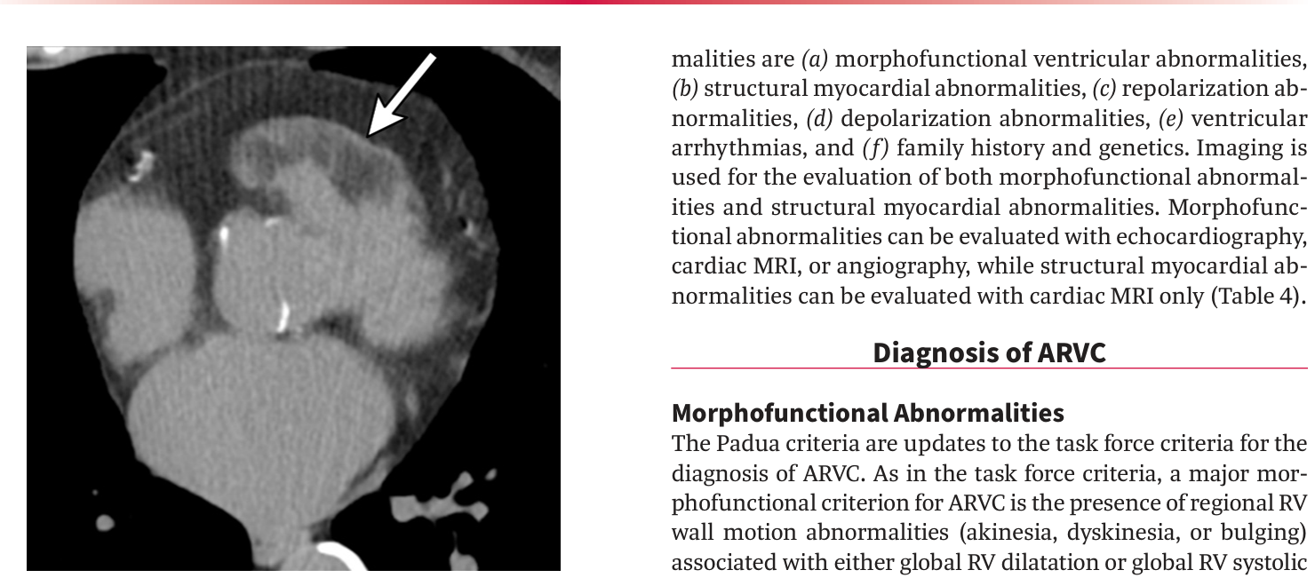
Figure 9. Figure 9. Physiologic presence of fat in the RV wall in an older woman. Axial non–contrast-enhanced CT image shows increased wall thickening (arrow) associated with myocardial fat infiltration, as opposed to ARVC, which is associated with wall thinning.解説: 高齢女性の右心室壁における生理的な脂肪の存在を示す、軸位造影なしCT画像です。心筋の脂肪浸潤を伴う壁厚の増加(矢印)が示されています。ARVCでは壁の菲薄化を伴うのに対し、この所見は異なります。
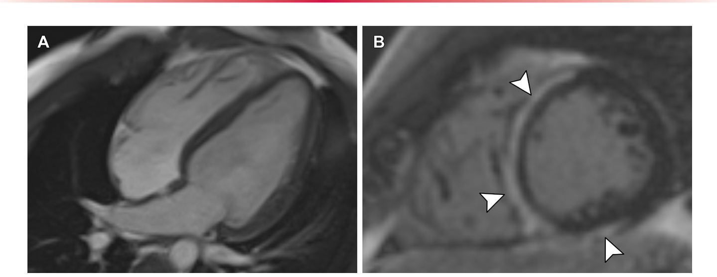
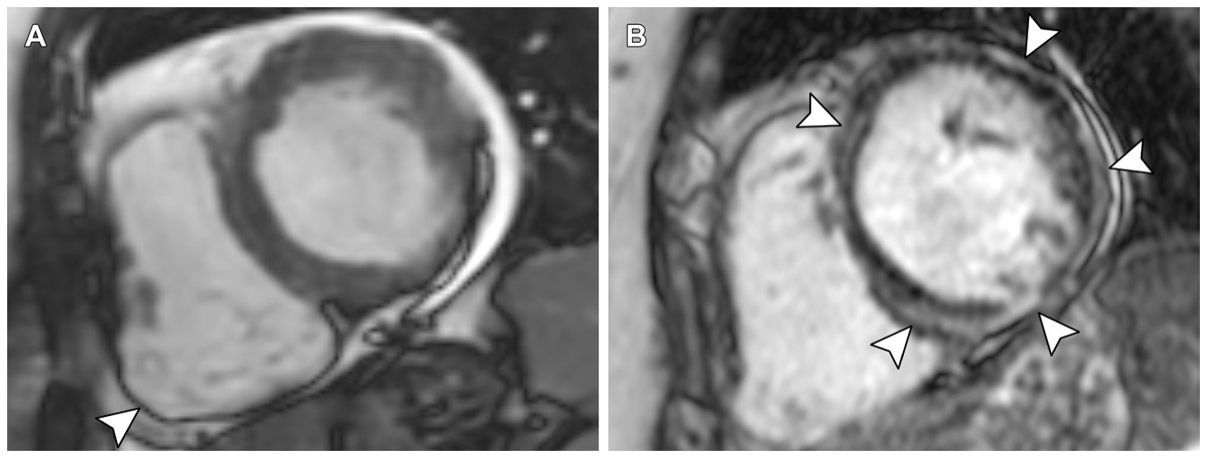
Figure 13. Figure 13. ALVC in an 18-year-old man with a DSP pathogenic variant who presented with ventricular tachycardia. (A) Still frame from cine SSFP cardiac MRI in a horizontal longitudinal axis plane shows mild LV enlargement with low-normal systolic function (ejection fraction, 54%). (B) LGE cardiac MR image in the short-axis plane at the midventricular level shows extensive abnormal delayed enhancement (arrowheads) in a subepicardial pattern involving the anterior, anteroseptal, inferoseptal, and inferior walls.
Figure 14
Figure 14. Figure 14. Biventricular ACM in a 63-year-old woman with a family history of ACM and a DSP pathogenic variant in a sister. (A) Still frame from cine SSFP cardiac MRI in the short-axis plane at end-systole shows a focal area of dyskinesia (arrowhead) in the RV free wall. The RV was normal in size (end-diastolic volume index, 75 mL/m2) and demonstrated reduced systolic function (ejection fraction, 36%), therefore fulfilling one major morphofunctional Padua criterion for RV involvement. The LV was also mildly dilated and showed depressed function (end-diastolic volume index, 108 mL/m2; ejection fraction, 36%) with regional hypokinesis, fulfilling two minor LV morphofunctional criteria. (B) LGE cardiac MR image in the short-axis plane at the midven- tricular level shows almost circumferential abnormal enhancement (arrowheads) in a subepicardial distribution, fulfilling one major Padua criterion for LV involvement and therefore suggesting the diagnosis of “definite biventricular ACM.” The patient was subsequently found to have a DSP variant.
Figure 15
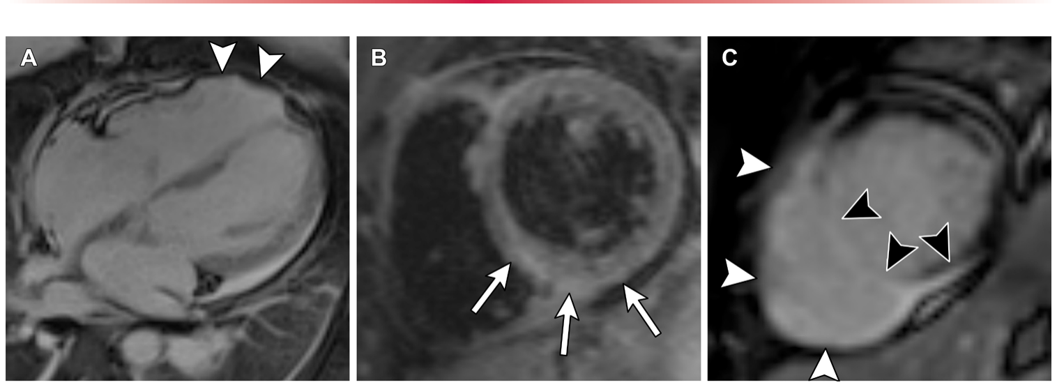
Figure 15. Figure 15. Biventricular ACM in a 26-year-old woman who presented with acute chest pain and presumed myocarditis. (A) Horizontal longitu- dinal axis (four-chamber) image from SSFP cine MRI shows a mildly dilated RV (119 mL/m2) and reduced function (ejection fraction, 31%) with a focal area of dyskinesis in the apical segment of the RV free wall (arrowheads) during systole. The left ventricular function was also depressed, with akinesis of the mid to apical septum. (B) Short-axis T2-weighted black-blood cardiac MR image at the mid ventricular level shows increased signal intensity of the LV myocardium (arrows) at the inferior and inferoseptal walls, consistent with edema. (C) Short-axis delayed-enhance- ment cardiac MR image at the apical level shows subepicardial LGE of the inferior wall, transmural LGE of the septal wall (black arrowheads), and circumferential LGE of the RV free wall (white arrowheads). Biopsy results were negative for giant cell myocarditis or other infiltrative diseases. Electrocardiography (not shown) demonstrated epsilon waves, and the patient had nonsustained ventricular tachycardia of different morphol- ogies. There was limited family history available because the patient was adopted. The clinical and MRI features fulfill the Padua criteria for biventricular ACM and the 2010 task force criteria for ARVC.
Figure 16
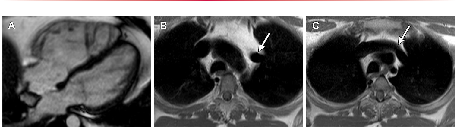
Figure 16. Figure 16. Partial anomalous pulmonary venous return in a 40-year-old woman with a history of palpitations and RV enlargement seen on an echocardiogram. (A) Still frame from cine SSFP cardiac MRI in a horizontal longitudinal plane shows a dilated RV (end-diastolic volume index, 211 mL/m2). (B, C) Axial T1-weighted fast spin-echo non–fat-suppressed MR images at the level of the aortic arch (B) and at the level of the origin of the arch vessels (C) show anomalous drainage of the left superior pulmonary vein into the left brachiocephalic vein (arrow), which was the cause of the RV enlargement.
Figure 18
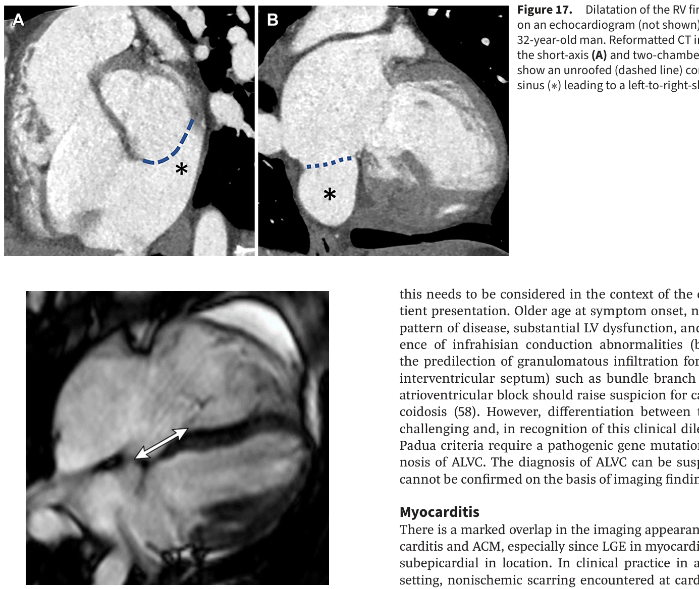
Figure 18. Figure 18. Dilated right ventricle seen previously on an echocardiogram (not shown) in a 17-year-old adolescent girl. Cardiac MR image acquired to evaluate for the pres- ence of ARVC shows a previously unsuspected Ebstein anomaly with apical displacement of the septal leaflet of the tricuspid valve (double-headed arrow).
Figure 20
Figure 20. Figure 20. Acute myocarditis in a 30-year-old man with a viral prodrome who presented in the emergency department with chest pain. LGE cardiac MR imag- es in the short-axis plane show subepicardial enhancement in the LV lateral wall (arrows). T2-weighted images (not shown) demonstrated high signal intensi- ty in the LV lateral wall.
Table 1
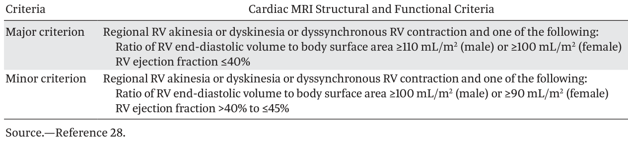
Table 1. Table 1: 2010 Task Force Criteria for the Diagnosis of ARVC with Cardiac MRI解説: 2010年タスクフォース基準に基づくARVCの心臓MRI構造機能基準の表です。主要基準として、右心室の局所無運動、異常運動、または非同期収縮に加えて、右心室拡張末期容積と体表面積の比が男性で110mL/m2以上、女性で100mL/m2以上、または右心室駆出率が40%以下であることが挙げられます。副次基準も同様の壁運動異常に加えて、右心室体積比が男性で100mL/m2以上、女性で90mL/m2以上、または駆出率が40%超45%以下の場合です。



コメントを残す