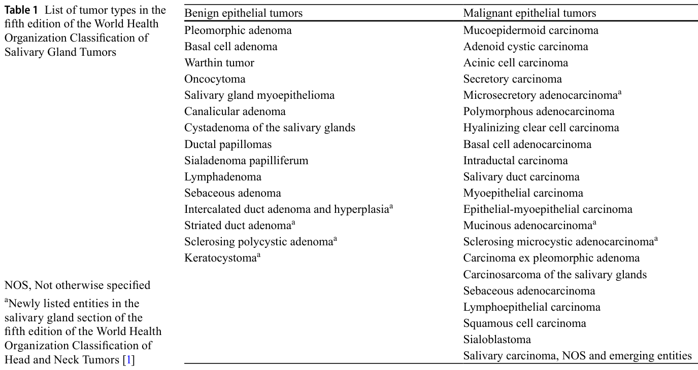
どうも、Beyond the Pixelです。唾液腺は、非常に多様な組織型の腫瘍が発生する場所であり、その診断は専門家にとっても複雑な課題となります。世界保健機関(WHO)による頭頸部腫瘍の分類は、第4版(2017年)から第5版へと改訂され、オンライン版が2022年3月に、WHOブルーブックが2024年3月に発行されました。この最新の分類では、唾液腺腫瘍(SGTs)は15の良性上皮性腫瘍と21の悪性上皮性腫瘍に分類されています。さらに、介在導管腺腫/過形成、線条導管腺腫、硬化性多嚢胞性腺腫、角化嚢胞腫の4つの新たな良性疾患が追加されました。
Table 1. Table 1 List of tumor types in the fifth edition of the World Health Organization Classification of Salivary Gland Tumors解説: 第5版WHO唾液腺腫瘍分類における腫瘍型の一覧です。良性上皮性腫瘍と悪性上皮性腫瘍がそれぞれ列挙されており、新たにリストされた疾患には注釈が付けられています。
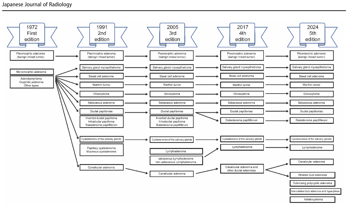
Figure 1. Fig. 1 Historical changes in the World Health Organization (WHO) classification of benign epithelial salivary gland tumors. The diagram illus trates the evolution of entities from the first edition (1972) through the current fifth edition (2024) (adapted from [32])解説: この図は、1972年の初版から2024年の第5版に至るまでの、世界保健機関(WHO)による良性上皮性唾液腺腫瘍の分類の歴史的変遷を示しています。
Table 2. Table 2 Summary of the clinicopathologic features of cystadenoma of the salivary glands, sebaceous adenoma, and lymphadenoma Feature Cystadenoma of the salivary glands Sebaceous adenoma Lymphadenoma Age and sex Infancy–91 years (median: 54 years) Slight female predominance解説: 唾液腺の嚢胞腺腫、脂腺腺腫、リンパ腺腫の臨床病理学的特徴をまとめたものです。年齢と性別、発生部位、臨床像、腫瘍の境界と被膜、肉眼像と大きさ、組織病理学と増殖パターン、細胞形態、間質、特徴的な免疫組織化学染色、腫瘍特異的遺伝子変異などの項目が含まれます。⚠ 自動抽出画像の検証で一致を確認できませんでした。正確な内容は元論文のTable 2をご参照ください。
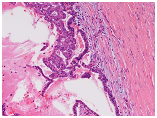
Figure 2. Fig. 2 Histopathology of cystadenoma of the salivary glands. High- power photomicrograph (hematoxylin and eosin [H&E] stain) shows a multicystic architecture with a cuboidal epithelial lining and intralu minal papillary projections解説: 唾液腺嚢胞腺腫の組織病理学的像を示す高倍率の顕微鏡写真(ヘマトキシリン・エオジン染色)です。多房性嚢胞性構造と立方状上皮性裏打ち、および管腔内乳頭状突起が観察されます。
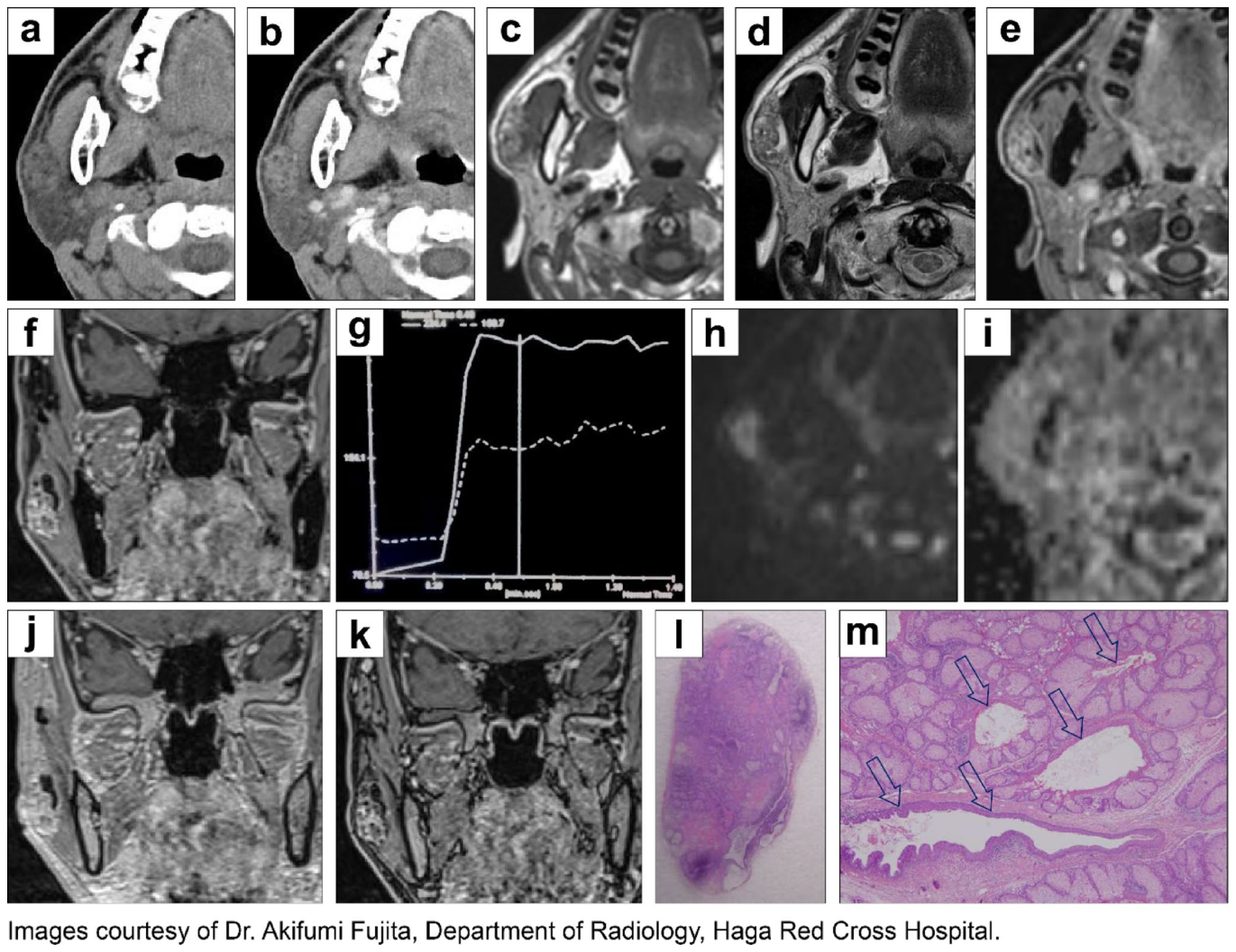
Figure 3. Fig. 3 Imaging and histopathology of sebaceous adenoma in a man in his 40s. a Non-contrast-enhanced computed tomography (CT) demon strates a soft-tissue-density mass with well-defined, smooth margins in the right parotid gland. Small, island-like low-density foci within the lesion correspond to fat. b Contrast-enhanced CT shows heteroge neous enhancement. c T1-weighted magnetic resonance (MR) imaging shows heterogeneous low-to-high signal intensity with small internal hyperintense foci. d T2-weighted MR imaging shows heterogeneous low-to-high signal intensity with small cyst-like and tubular hyper intense structures. e, f Axial (e) and coronal (f) gadolinium-enhanced T1-weighted MR imaging demonstrates heterogeneous enhancement. g Time–signal intensity curve on dynamic contrast-enhanced (DCE) MR imaging shows a rapid enhancement followed by a plateau pat解説: 40歳代男性の脂腺腺腫の画像と組織病理学的像です。(a)非造影CTでは、右耳下腺に境界明瞭で平滑な辺縁を持つ軟部組織密度の腫瘤が示されています。(b)造影CTでは不均一な増強効果が見られます。(c)T1強調MR画像では不均一な低~高信号を示し、(d)T2強調MR画像では小さな嚢胞様および管状の過信号構造が確認されます。(j) in-phaseと(k) opposed-phase化学シフトMR画像では、病変内の焦点性信号低下が観察され、微細な脂肪の存在が確認されます。(l)は薄い線維性被膜に囲まれた境界明瞭な腫瘍を示し、(m)は脂腺分化を伴う上皮細胞の増殖が観察される高倍率の顕微鏡写真です。
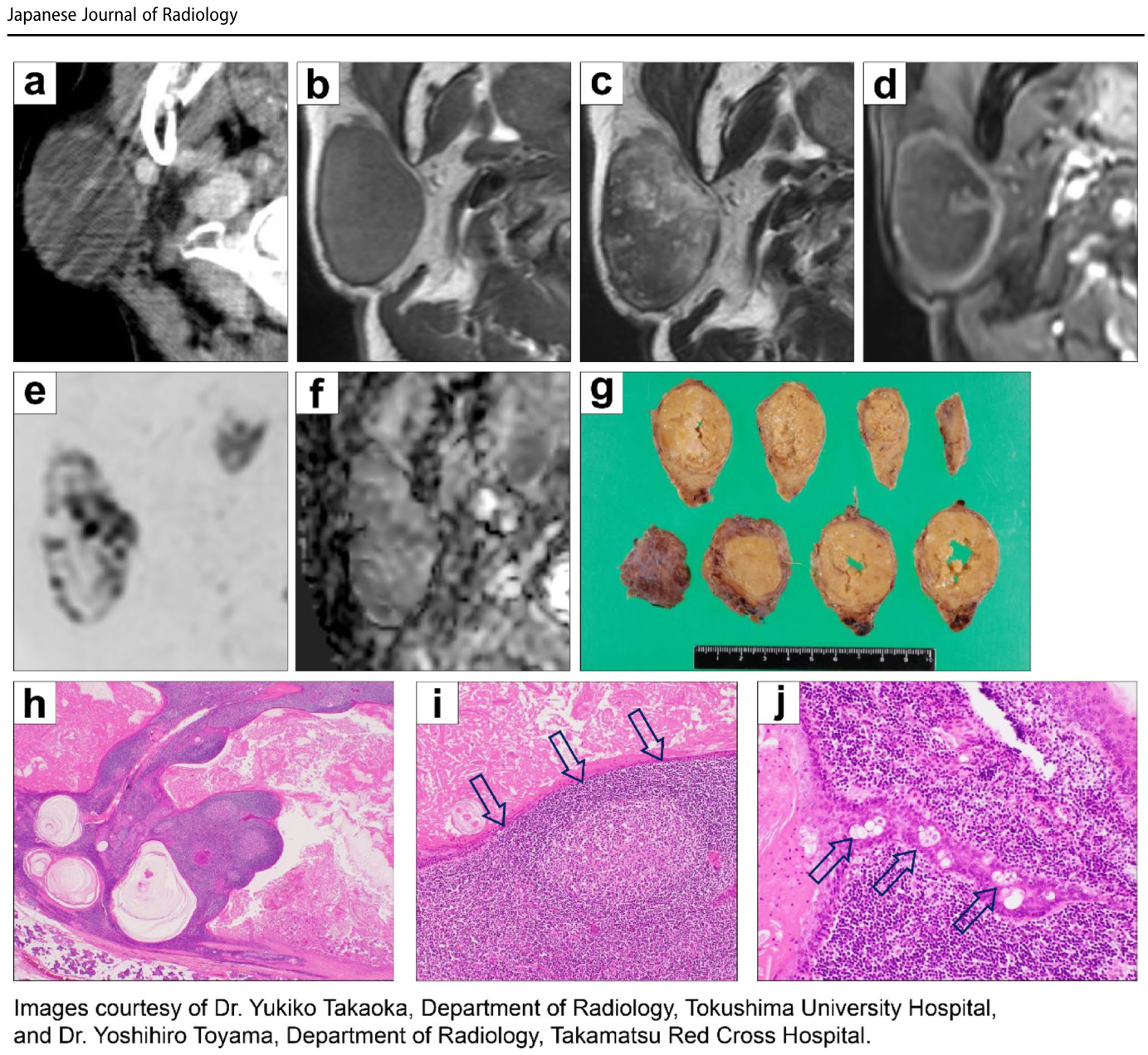
Figure 4. Fig. 4 Imaging and histopathology of sebaceous lymphadenoma in a woman in her 60s. a Contrast-enhanced computed tomography (CT) demonstrates a mass with well-defined smooth margins in the right parotid gland. Thick capsular and septal enhancement is seen; however, the majority of the lesion shows poor enhancement. b T1-weighted magnetic resonance (MR) imaging shows low signal intensity with a prominent low-signal rim corresponding to the fibrous capsule. c T2-weighted MR imaging shows predominantly low signal intensity with focal hyperintense areas, resulting in a heterogeneous appear ance. d Contrast-enhanced T1-weighted MR imaging confirms thick capsular and septal enhancement with poor central enhancement. e, f Axial diffusion-weighted whole-body imaging with background body signal suppression, displayed with inverted grayscale contrast (e), and解説: 60歳代女性の脂腺リンパ腺腫の画像と組織病理学的像です。(a)造影CTでは右耳下腺に境界明瞭な腫瘤が見られ、厚い被膜と隔壁の増強効果が示されていますが、病変の大部分は乏しい増強効果を示しています。(b)T1強調MR画像では低信号を示し、(c)T2強調MR画像では主に低信号ですが、焦点性の高信号領域が混在し不均一な外観を呈しています。(d)造影T1強調MR画像では厚い被膜と隔壁の増強効果が確認されます。(g)は50x50x34mmの黄色い腫瘤で、嚢胞性スペースには脂腺性残骸が含まれています。(h)は境界明瞭で線維性被膜に囲まれた腫瘍を示し、(i)は反応性リンパ様間質と胚中心を示しています。(j)はリンパ様間質内に埋め込まれた腫瘍細胞索状構造を示し、散在する脂腺分化を示す細胞が観察されます。
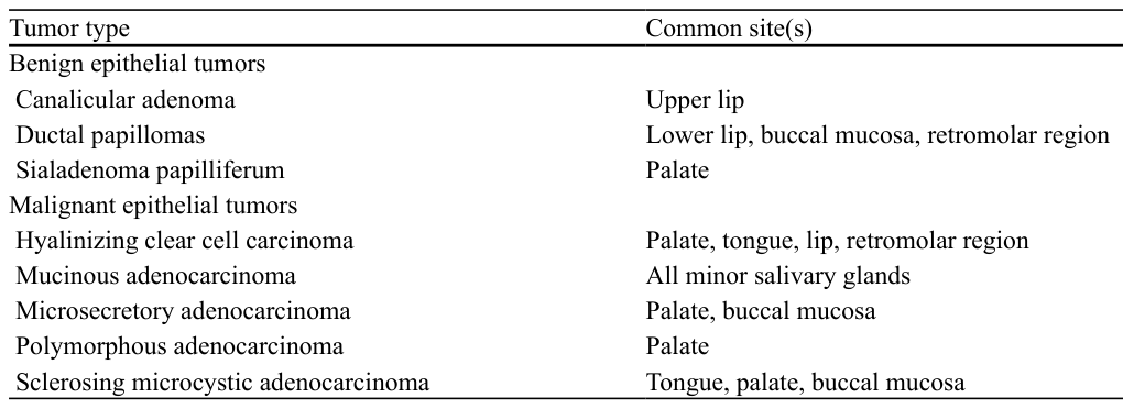
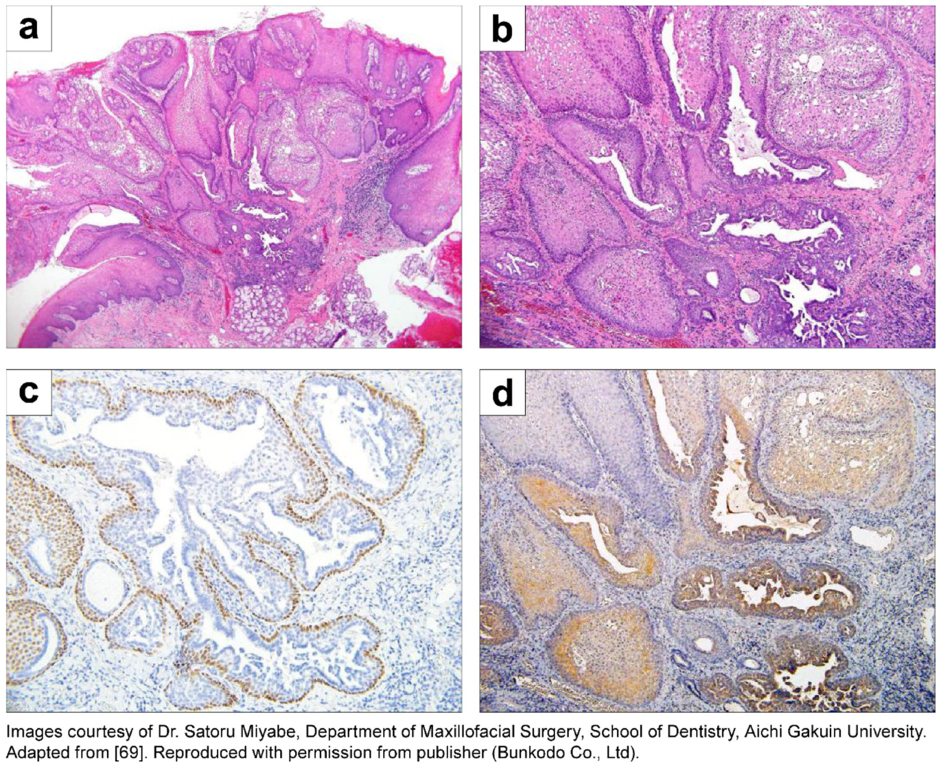
Table 3. Table 3 Tumor types that occur predominantly in intraoral minor salivary glands and their common sites [69]解説: 口腔内小唾液腺に主に発生する腫瘍型とその一般的な発生部位を示しています。良性上皮性腫瘍と悪性上皮性腫瘍の両方が含まれています。
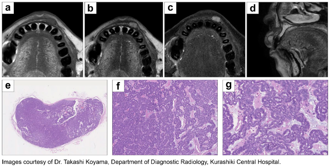
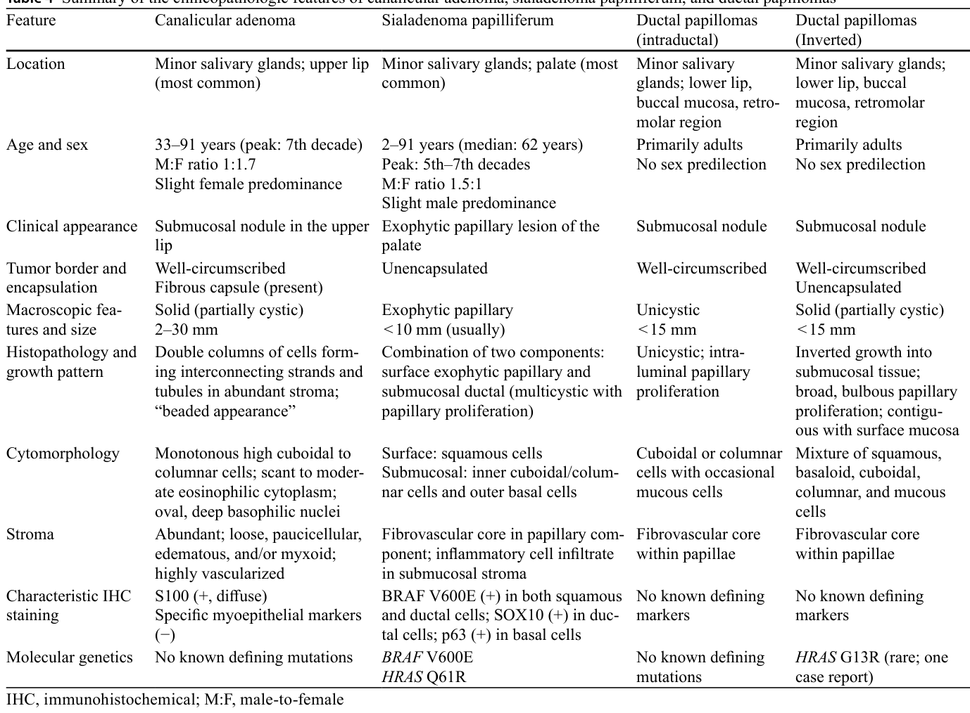
Figure 5. Fig. 5 Imaging and histopathology of canalicular adenoma in a woman in her 70s. a–d Axial T1-weighted (a), axial T2-weighted (b), axial fat-suppressed T2-weighted (c), and sagittal T2-weighted (d) mag netic resonance (MR) imaging demonstrates a well-defined mass in the upper lip. The lesion is homogeneously hypointense on T1-weighted imaging and heterogeneously hyperintense on T2-weighted imaging. e Low-power photomicrograph (hematoxylin and eosin [H&E] stain)解説: 70歳代女性の上唇に発生した細管腺腫の画像と組織病理学的像です。(a)軸位T1強調、(b)軸位T2強調、(c)軸位脂肪抑制T2強調、(d)矢状T2強調MR画像は、上唇に境界明瞭な腫瘤を示しています。病変はT1強調画像で均一な低信号、T2強調画像で不均一な高信号を示します。(e)低倍率の顕微鏡写真では、淡い好塩基性を示す境界明瞭な腫瘍が確認されます。(f)中倍率および(g)高倍率の顕微鏡写真では、単層の単調な円柱状上皮細胞が特徴的な細管状(「ビーズ状のネックレス」)パターンで配置されていることが示されています。
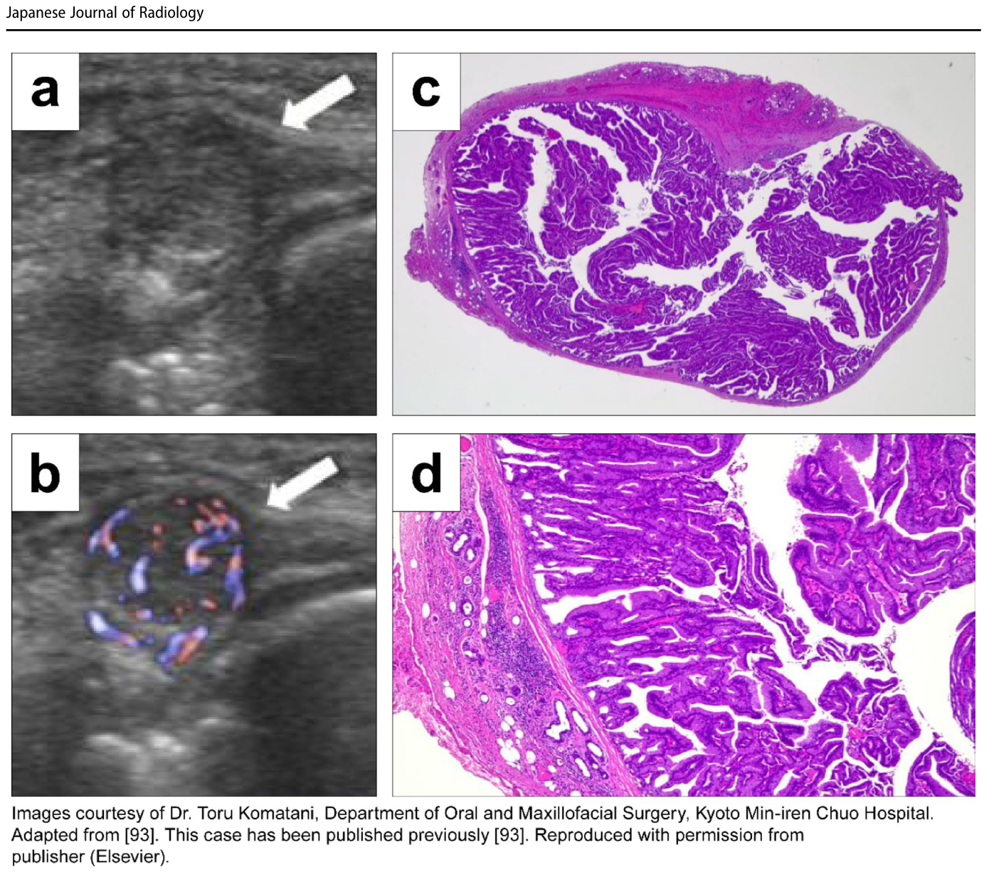
Figure 7. Fig. 7 Imaging and histopathology of intraductal papilloma in a woman in her 40s. Adapted from [93]. a Ultrasonography demon strates a cystic mass containing internal echogenic material (arrow). b Color Doppler ultrasonography shows internal vascular flow (arrow). c Low-power photomicrograph (hematoxylin and eosin [H&E] stain)解説: 40歳代女性の管内乳頭腫の画像と組織病理学的像です。(a)超音波検査では、内部エコーを伴う嚢胞性腫瘤が示されています。(b)カラードップラー超音波では内部血流が確認されます。(c)低倍率の顕微鏡写真(ヘマトキシリン・エオジン染色)では、乳頭状上皮増殖で満たされた境界明瞭な単房性嚢胞性腫瘤が示されています。(d)高倍率の顕微鏡写真では、良性な円柱状上皮細胞に裏打ちされた薄い線維血管性中心を持つ乳頭が示されています。


コメントを残す